 MSK Academy
1. Ventral view
1. Ventral view
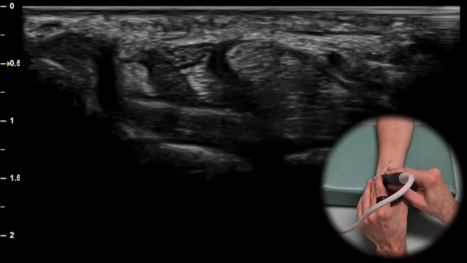
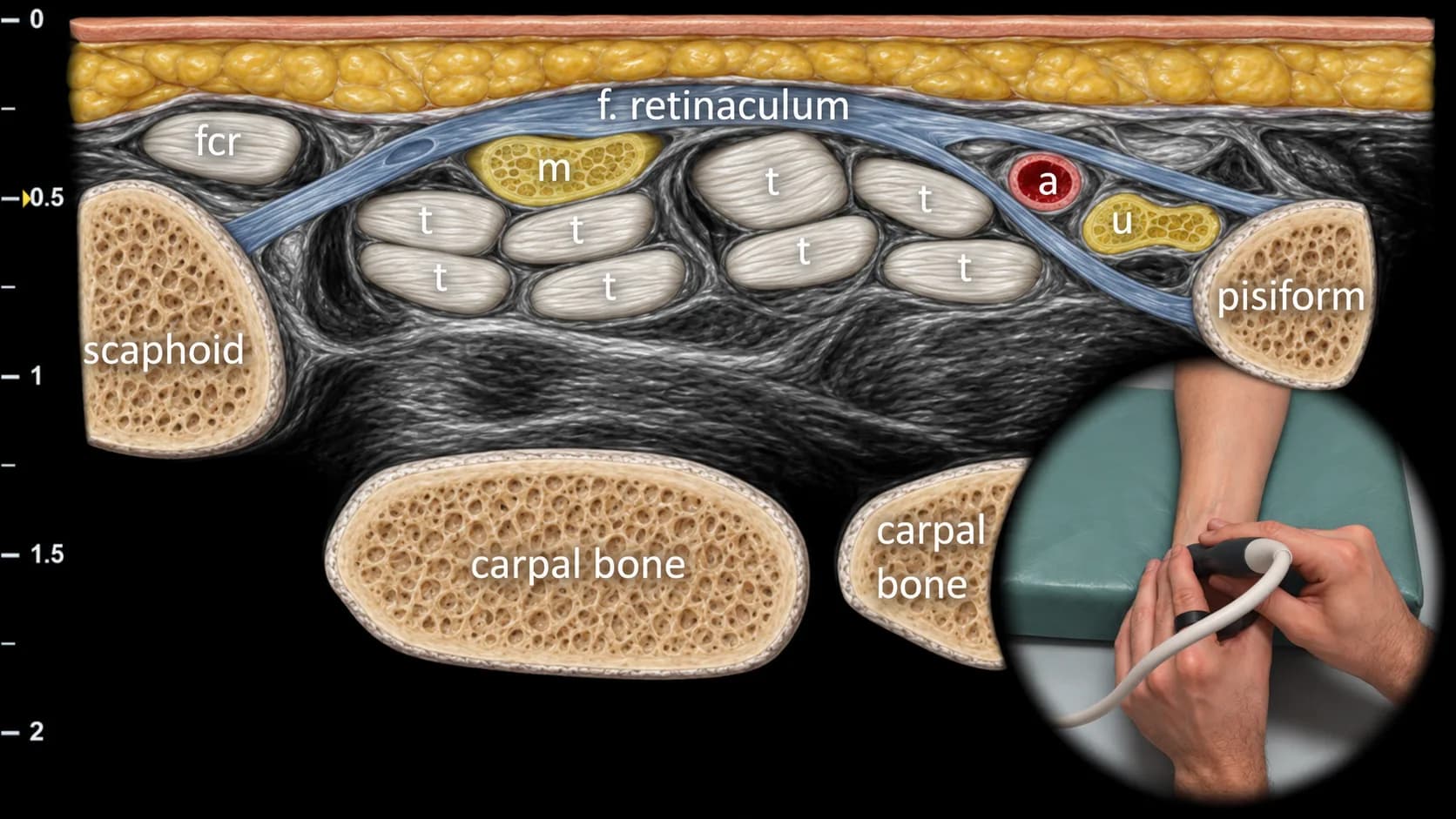
Figure 1. Ventral view, transverse plane. fcr: m. flexor carpi radialis, m: n. medianus, a: a. ulnaris, u: n. ulnaris, t: flexor tendons
Transverse ultrasound section of the volar side of the wrist at the level of the carpal tunnel and Guyon's canal. Key bony landmarks include the os scaphoideum radially and os pisiforme ulnarly, between which the flexor retinaculum can be seen overlying the flexor tendons (t) and n. medianus (m) in the carpal tunnel region. Ulnar to this area, the a. ulnaris (a) and n. ulnaris (u) are visible as they pass through Guyon's canal, while radially the tendon of m. flexor carpi radialis (fcr) is shown. This projection is essential for evaluating compressive neuropathies of the n. medianus and n. ulnaris, as well as for detecting flexor tenosynovitis and the presence of ganglion cysts in the volar wrist region.
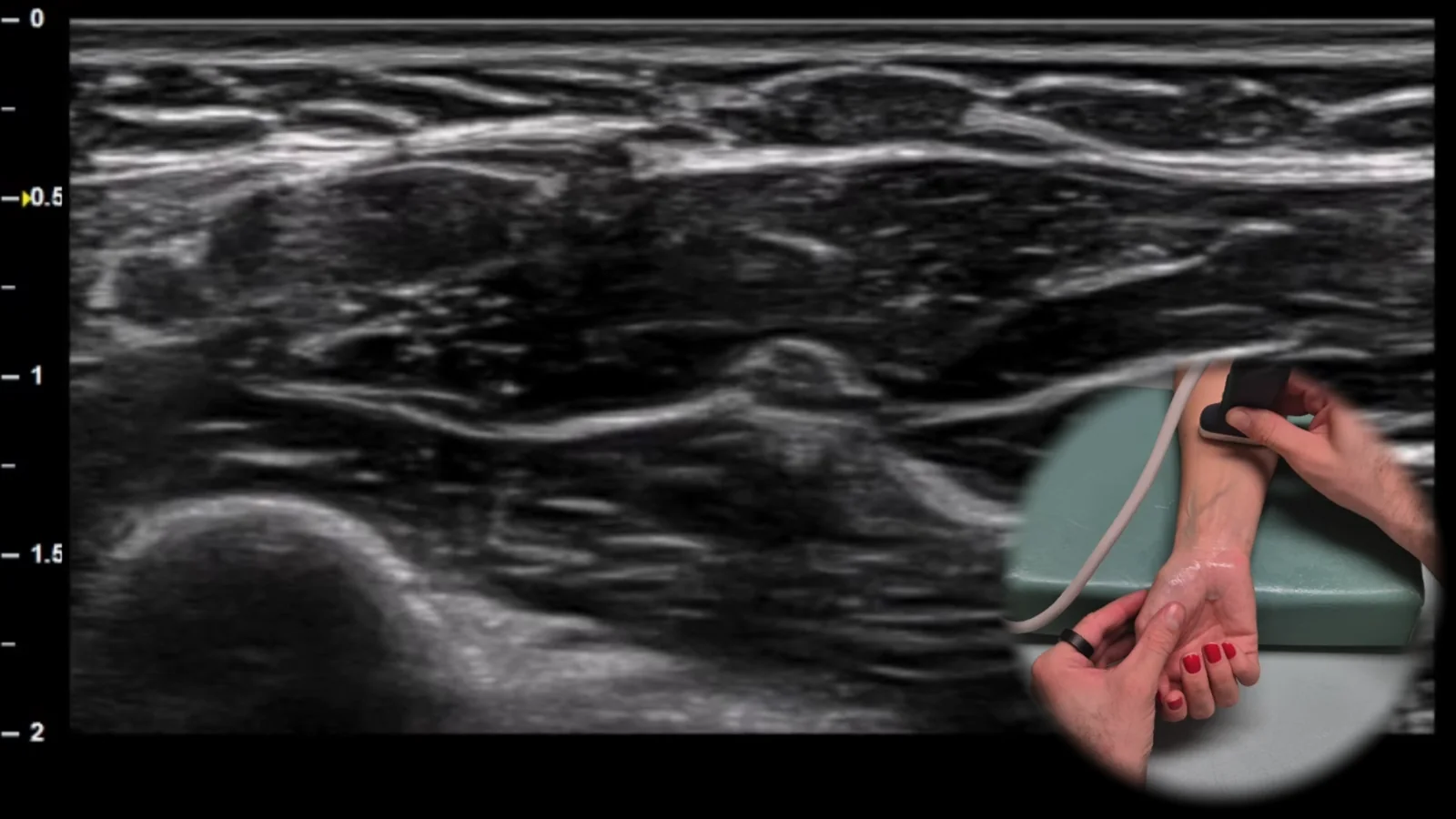
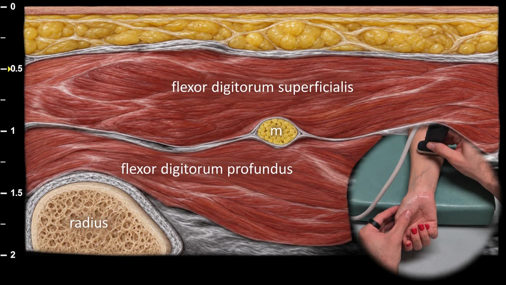
Figure 2. Ventral view, transverse plane. m: median nerve
Transverse ultrasound section of the volar aspect of the distal forearm during proximal tracking of the median nerve from the carpal tunnel area. The median nerve (m) is positioned in this projection between the flexor digitorum superficialis and flexor digitorum profundus muscles, with the radius visible deeper as a bony landmark. At this level, the median nerve has a typical fascicular ("honeycomb") appearance, which is important for assessing its morphology and for detecting pathological changes in compressive neuropathies or other nerve involvement.
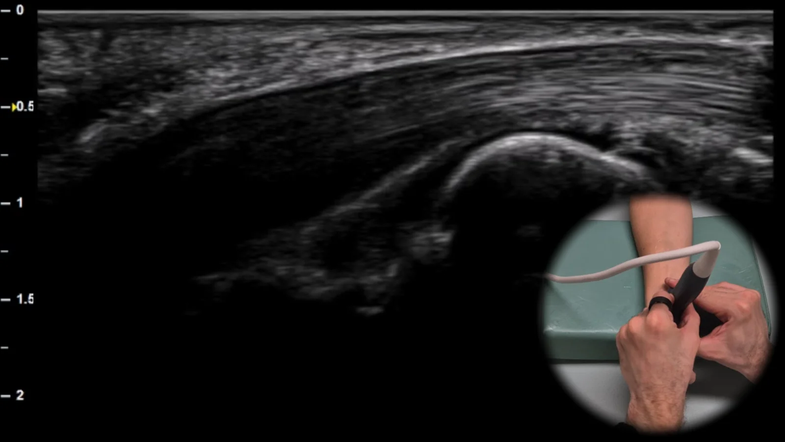
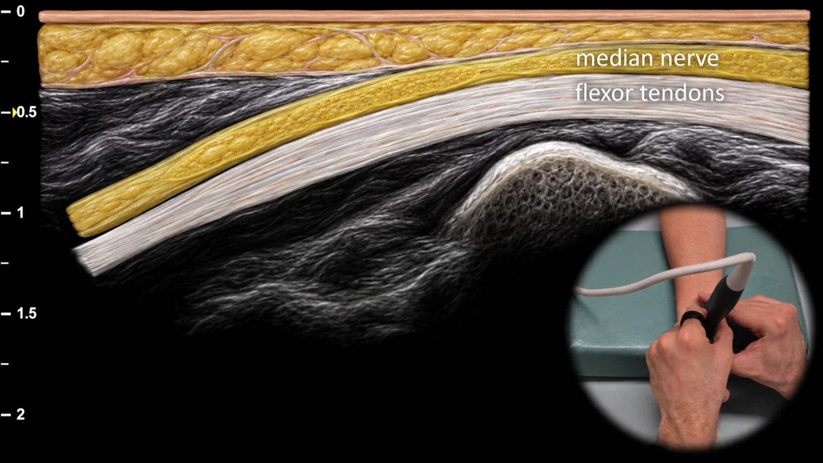
Figure 3. Ventral view, sagittal plane. m: n. medianus
Longitudinal ultrasound section of the volar side of the wrist showing the n. medianus at the entrance to the carpal tunnel. The nervus medianus (m) is visible in this projection as a striated structure with parallel echogenic lines, located superficially above the flexor tendons, which corresponds to its typical longitudinal fascicular architecture. This projection is important for assessing nerve continuity, changes in its course, and potential signs of compression at the carpal tunnel entrance.
Clinical Note
In median nerve compression, focal narrowing of the nerve with proximal dilatation may be visible at the site of entry into the carpal tunnel, which in longitudinal view resembles a "bottle neck" shape.
Access the full Health Library
Full access to scanning protocols, anatomy, and clinical references. Cancel anytime.
- Every protocol and anatomy reference
- Original ultrasound illustrations and video demonstrations
- Sync across mobile and web