 MSK Academy
2. Dorsale Ansicht
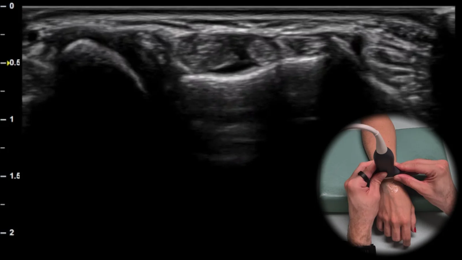
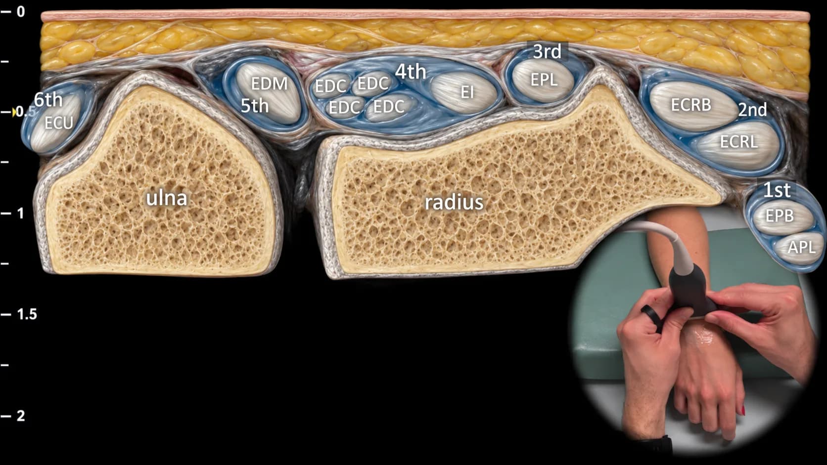
Abbildung 1. Dorsale Ansicht, Transversalebene. ECU: m. extensor carpi ulnaris, EDM: m. extensor digiti minimi, EDC: m. extensor digitorum communis, EI: m. extensor indicis, EPL: m. extensor pollicis longus, ECRB: m. extensor carpi radialis brevis, ECRL: m. extensor carpi radialis longus, EPB: m. extensor pollicis brevis, APL: m. abductor pollicis longus
Sonografischer Querschnitt durch die dorsale Seite des Handgelenks auf Höhe des distalen Radius. In dieser Projektion lassen sich alle sechs Strecksehnenfächer klar darstellen, von der ulnar gelegenen Sehne des m. extensor carpi ulnaris bis zu den radial gelegenen Sehnen des ersten Strecksehnenfachs, insbesondere des m. abductor pollicis longus und des m. extensor pollicis brevis; dazwischen sind auch die über den distalen Radius und die distale Ulna verlaufenden Sehnen der anderen Extensoren sichtbar. Diese Projektion ist für die orientierende Beurteilung der Strecksehnenfächer und für den Nachweis von Tenosynovitiden, Subluxationen und degenerativen Veränderungen der Sehnen im dorsalen Handgelenksbereich unerlässlich.
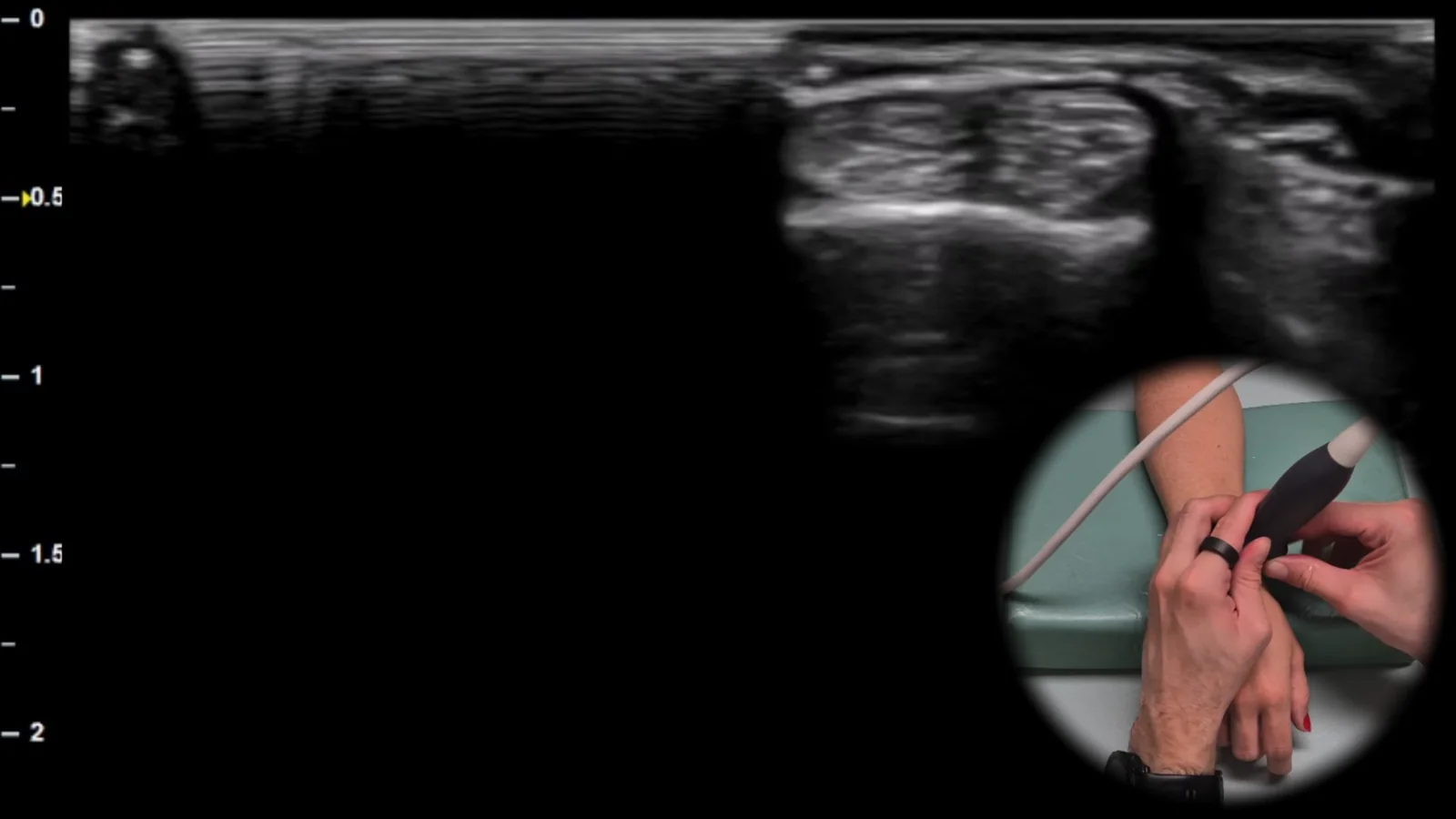
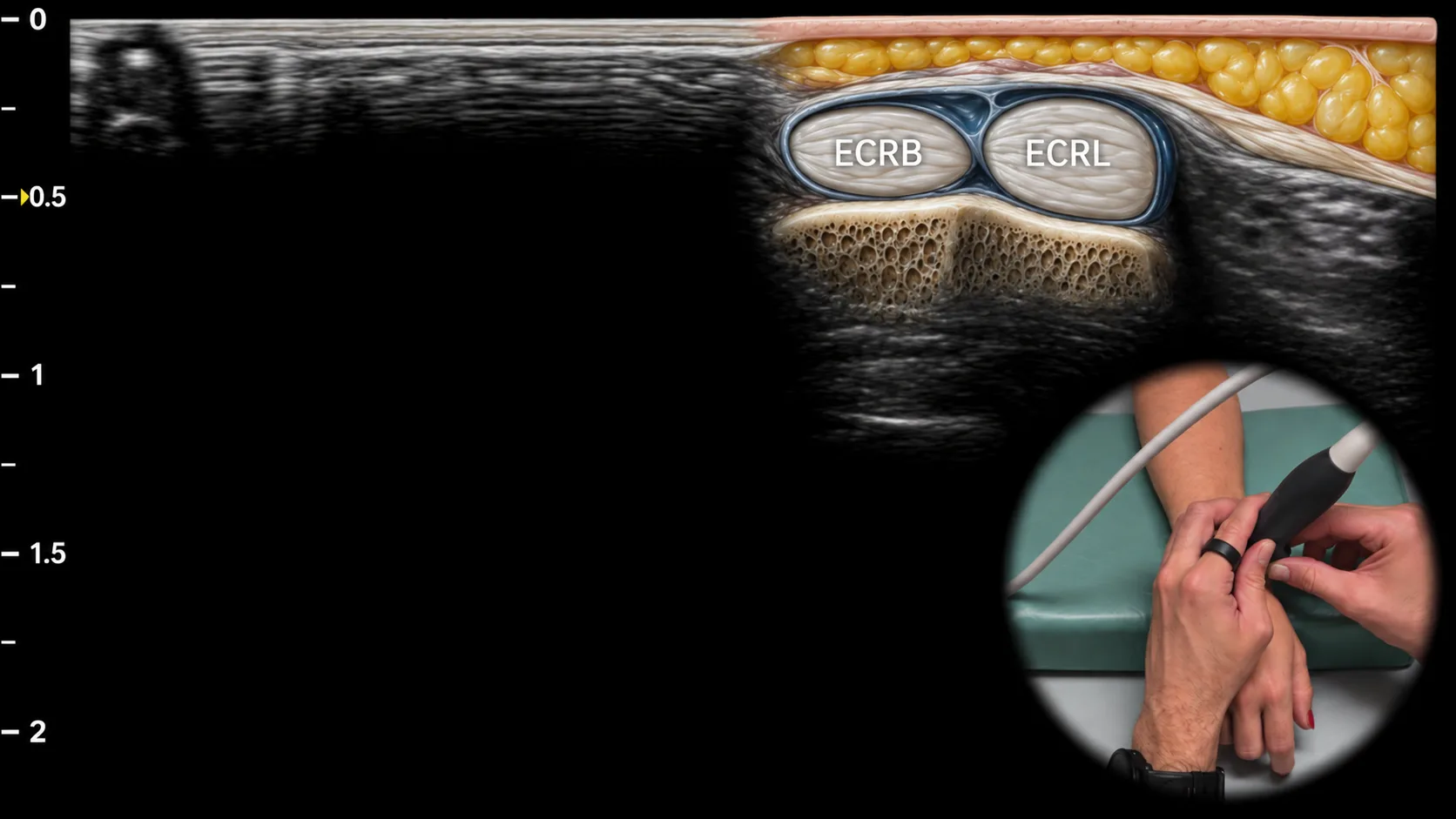
Abbildung 2. Dorsalansicht, Transversalebene. ECRB: m. extensor carpi radialis brevis, ECRL: m. extensor carpi radialis longus
Transversaler Ultraschallschnitt durch den dorsoradialen Anteil des Handgelenks mit Darstellung des zweiten Strecksehnenfachs, das lateral des Lister-Tuberkels liegt. In dieser Ansicht sind die Sehnen des m. extensor carpi radialis brevis (ECRB) und des m. extensor carpi radialis longus (ECRL) sichtbar, die nahe der dorsalen Kontur des distalen Radius verlaufen. Dieser Bereich ist häufig von Überlastung betroffen, und diese Ansicht ist wichtig für die Beurteilung von Tenosynovitis, irritativen Veränderungen und anderen pathologischen Veränderungen der Sehnen des zweiten Strecksehnenfachs.
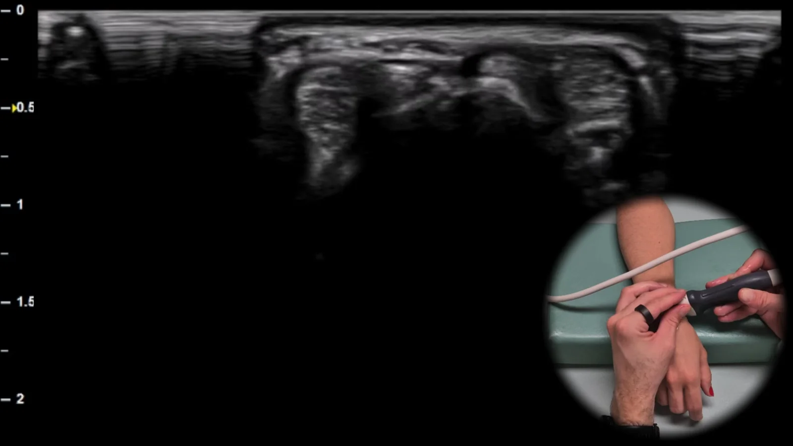
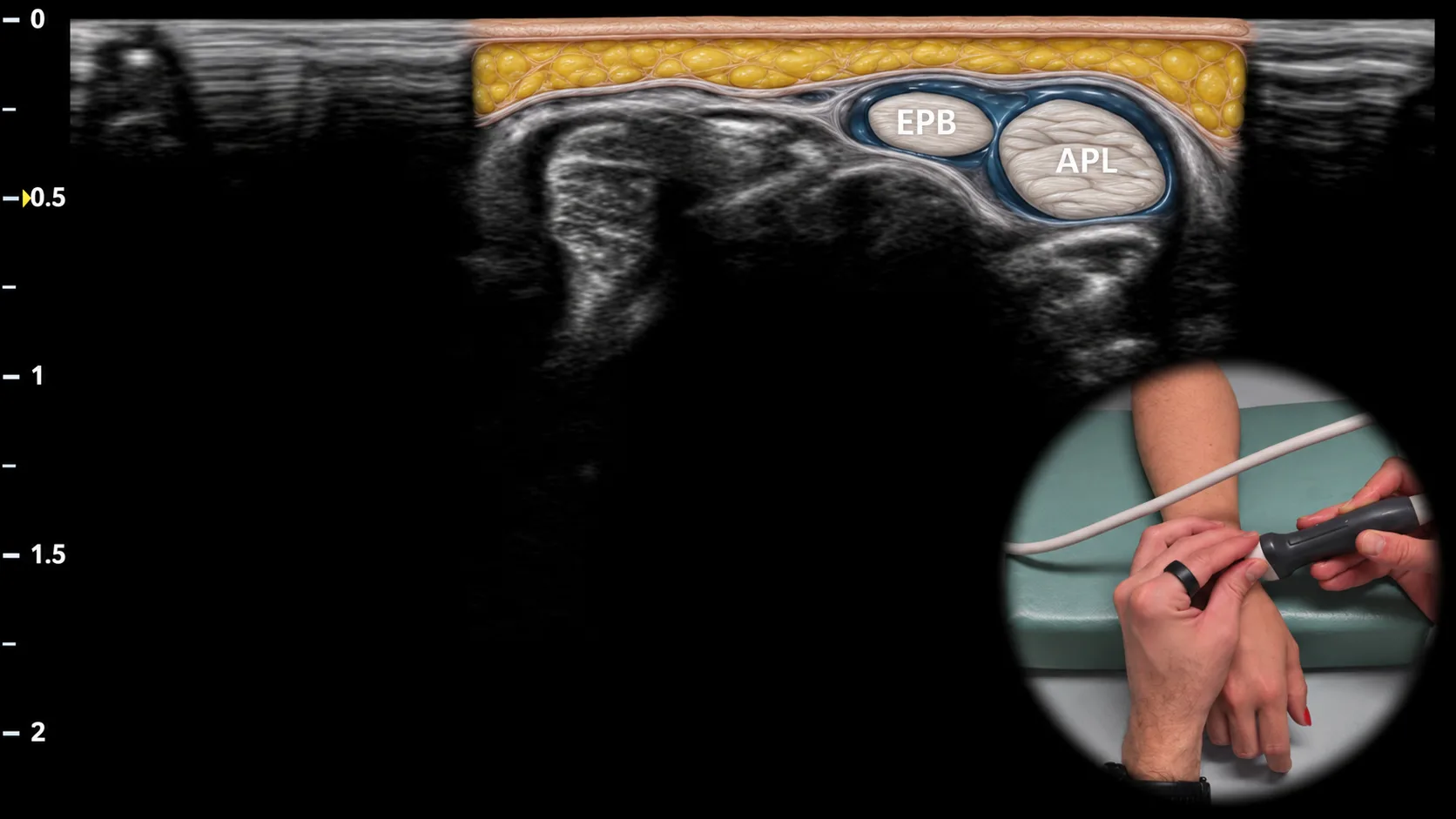
Abbildung 3. Dorsale Ansicht, Transversalebene. EPB: m. extensor pollicis brevis, APL: m. abductor pollicis longus
Transversaler sonografischer Schnitt durch den dorsoradialen Teil des Handgelenks mit Darstellung des ersten Strecksehnenfachs. In dieser Projektion sind die Sehnen des m. abductor pollicis longus (APL) und des m. extensor pollicis brevis (EPB) sichtbar, die in einer gemeinsamen Sehnenscheide über dem Processus styloideus radii verlaufen. Diese Projektion ist typisch für die Beurteilung der De-Quervain-Tenosynovitis und ermöglicht die Beurteilung einer Verdickung der Sehnenscheide, des Vorhandenseins von Flüssigkeit und weiterer Anzeichen einer Überlastung im Bereich des ersten Strecksehnenfachs.
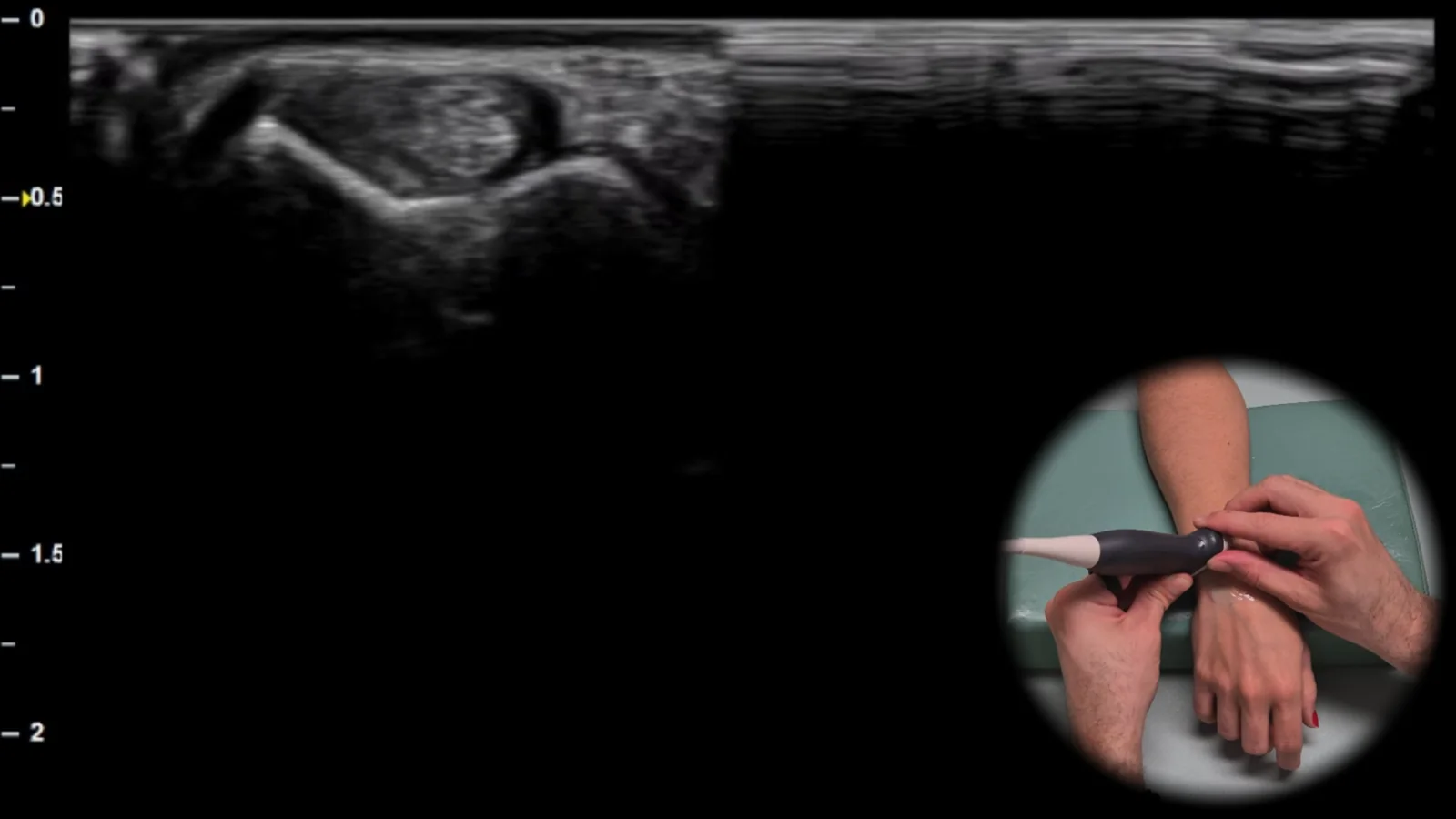
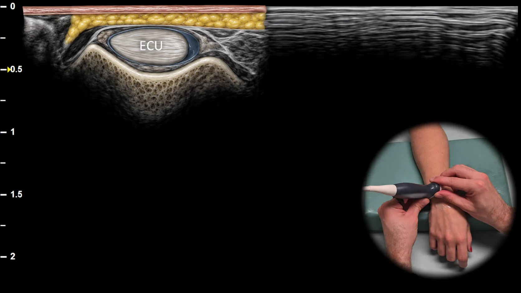
Abbildung 4 Dorsalansicht, Transversalebene. ECU: M. extensor carpi ulnaris
Transversaler Ultraschallschnitt durch den dorsoulnaren Anteil des Handgelenks mit Darstellung des über der Ulna gelegenen sechsten Strecksehnenfachs. In dieser Projektion ist die Sehne des M. extensor carpi ulnaris (ECU) sichtbar, die als Hauptstruktur dieses Strecksehnenfachs in der Rinne an der Ulna verläuft. Diese Projektion ist für die Beurteilung von Tenosynovitis, Instabilität und Subluxation der ECU-Sehne von wesentlicher Bedeutung, insbesondere bei der dynamischen Untersuchung im Zusammenhang mit ulnaren Handgelenkschmerzen.
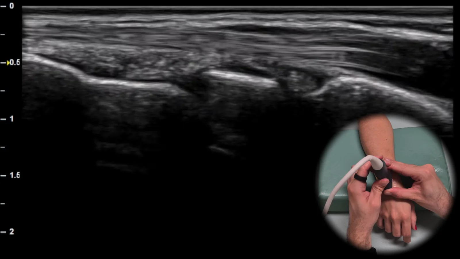
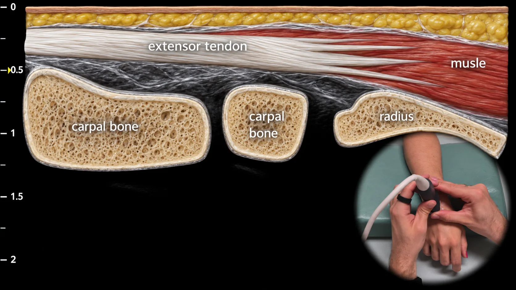
Abbildung 5. Dorsalansicht, Sagittalebene.
Sonografischer Längsschnitt der Dorsalseite des Handgelenks mit Darstellung der Extensorsehnen in der Längsachse über dem Radiokarpalgelenk. In dieser Projektion sind die Extensorsehnen als fibrilläre Strukturen sichtbar, die oberflächlich über der Kontur des distalen Radius und der Handwurzelknochen verlaufen; die Bildgebung schließt auch den Bereich des Radiokarpalgelenks ein. Diese Projektion ist wichtig zum Nachweis eines Gelenkergusses und von Zeichen einer Synovitis sowie zur Beurteilung der Kontinuität der Extensorsehnen bei Verdacht auf eine partielle oder vollständige Sehnenverletzung.
Zugang zur gesamten Bibliothek
Voller Zugriff auf Scan-Protokolle, Anatomie und klinische Referenzen. Jederzeit kündbar.
- Alle Protokolle und Anatomiereferenzen
- Originale Ultraschall-Illustrationen und Video-Demonstrationen
- Synchronisierung zwischen Mobilgerät und Web