 MSK Academy
4. Dorsal view
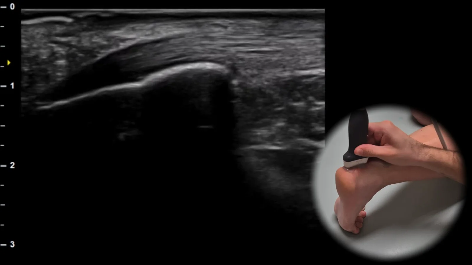
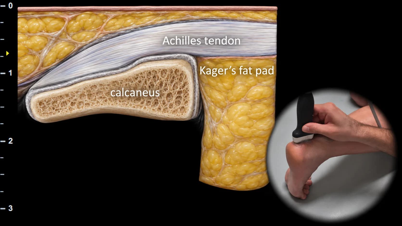
Figure 1. Dorsal view, sagittal plane.
Longitudinal ultrasound section of the posterior ankle showing the Achilles tendon as a thick hyperechoic fibrillar band running to its insertion on the calcaneus. Below the tendon, Kager's fat pad is visible, which forms an important anatomical landmark in the posterior ankle and contributes to clear visualization of the relationship between the Achilles tendon and the calcaneus. This projection is essential for assessing Achilles tendon continuity, detecting tendinopathy, partial ruptures, and signs of retrocalcaneal bursitis or changes in surrounding soft tissues.
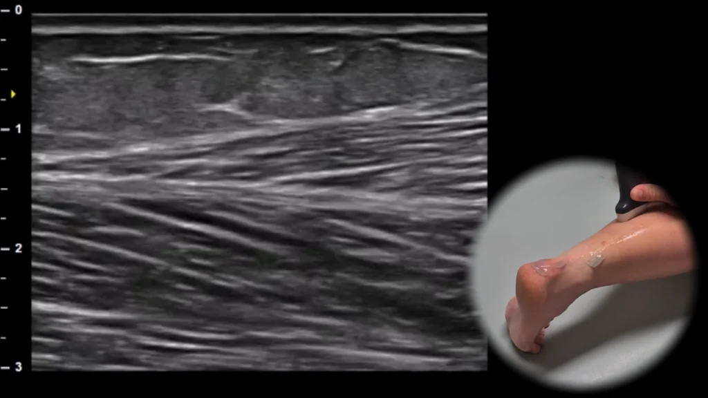
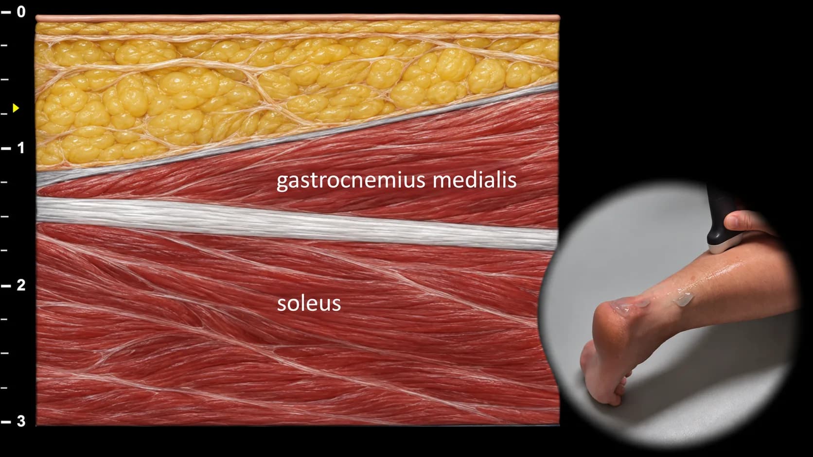
Figure 2. Dorsal view, sagittal plane.
Longitudinal ultrasound section of the posterior calf showing the myotendinous interface of m. gastrocnemius medialis and m. soleus. The m. gastrocnemius medialis is visible superficially, m. soleus deeper, and between them an aponeurotic line, which forms an important landmark of this projection. This projection is essential for evaluating the myotendinous junction of m. triceps surae and for detecting muscle ruptures, hematomas, or other lesions in the area of the medial head of m. gastrocnemius and m. soleus.
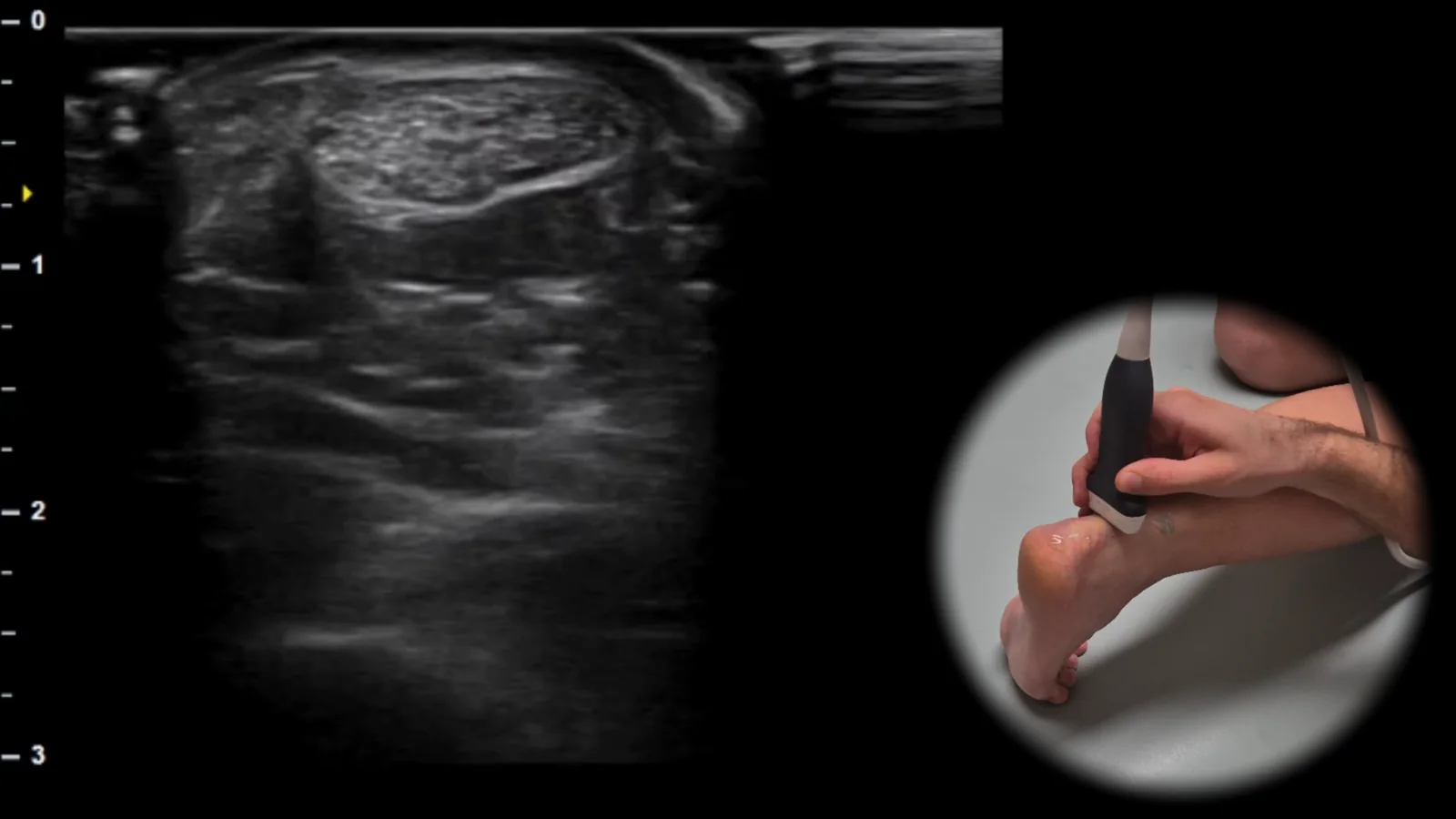
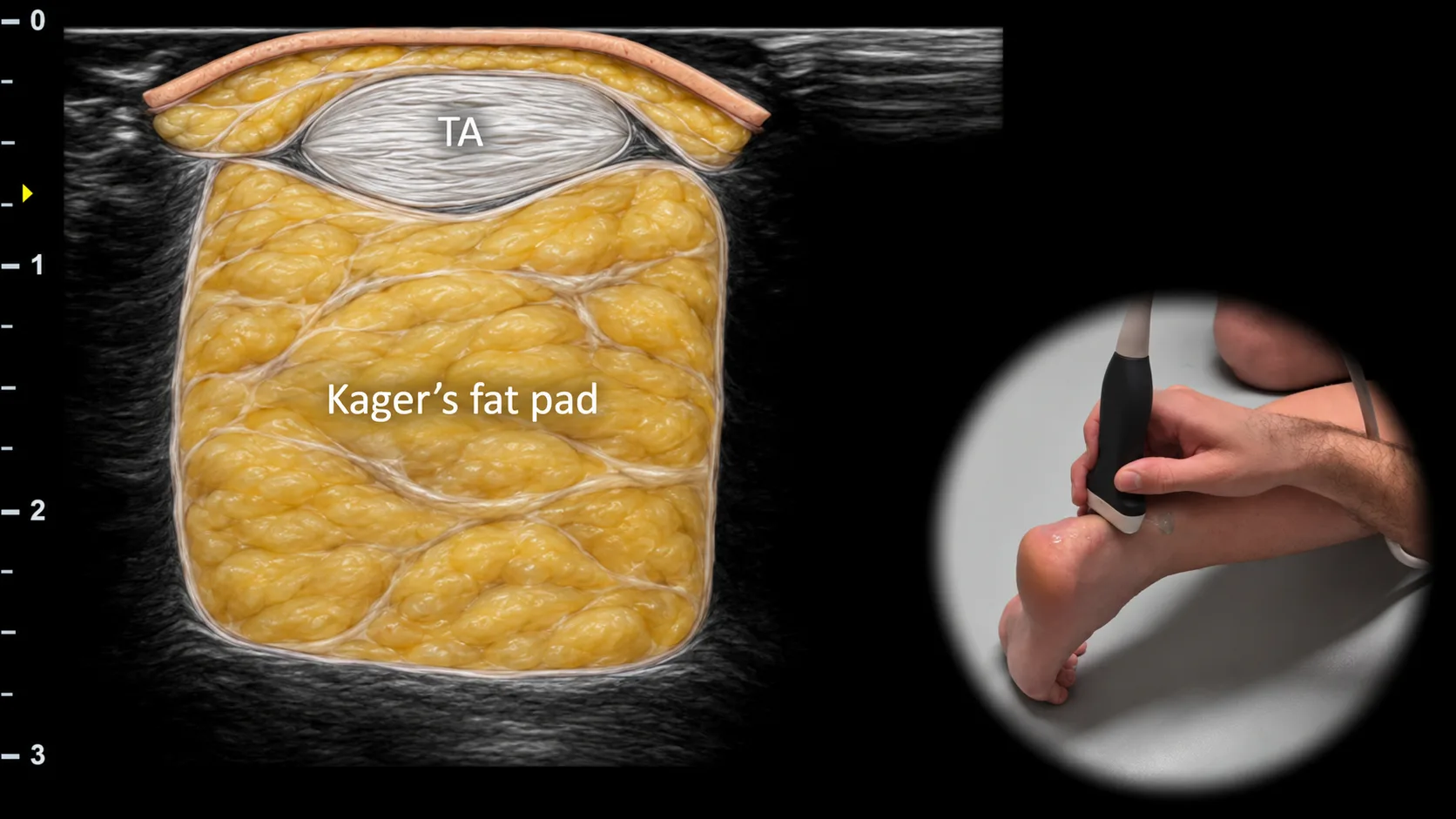
Figure 3. Dorsal view, transverse plane. TA: Achilles tendon
Transverse ultrasound section of the posterior ankle showing the Achilles tendon (TA) as an oval fibrillar structure located superficially above Kager's fat pad. Kager's fat pad forms a homogeneous soft tissue space ventral to the Achilles tendon and represents an important landmark for this projection. This projection is essential for evaluating the thickness, echostructure and continuity of the Achilles tendon and for detecting tendinopathy, partial ruptures or changes in surrounding soft tissues.
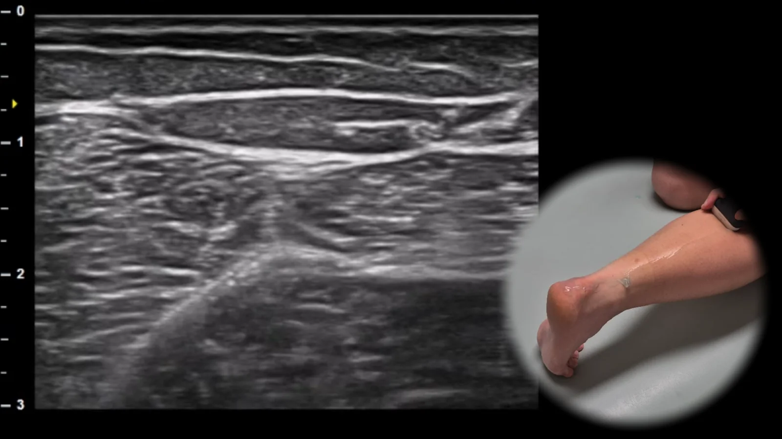
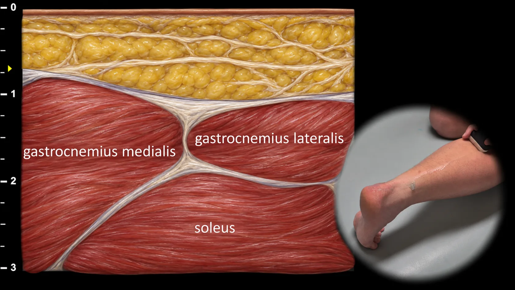
Figure 4. Dorsal view, transverse plane.
Transverse ultrasound section of the posterior part of the leg showing the muscle group m. triceps surae. Superficially, the medial and lateral heads of m. gastrocnemius are visible, between which the deeper m. soleus lies, forming the floor of this projection. This arrangement allows for clear visualization of the mutual anatomical relationships of the individual parts of triceps surae. This projection is essential for evaluating muscle architecture, detecting myotendinous injuries, and assessing chronic changes such as atrophy or fibrosis.
Access the full Health Library
Full access to scanning protocols, anatomy, and clinical references. Cancel anytime.
- Every protocol and anatomy reference
- Original ultrasound illustrations and video demonstrations
- Sync across mobile and web