 MSK Academy
1. Ventral view
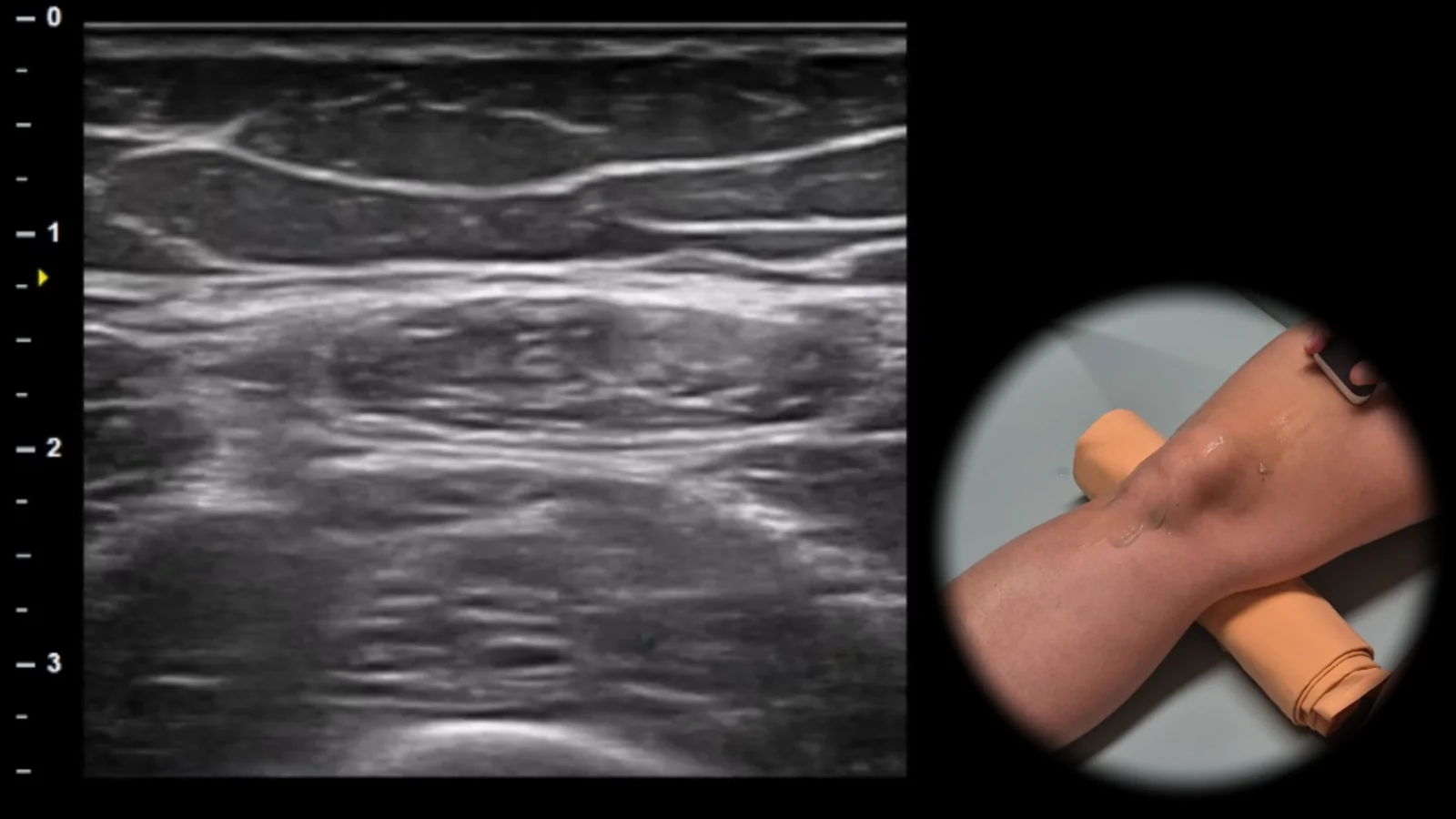
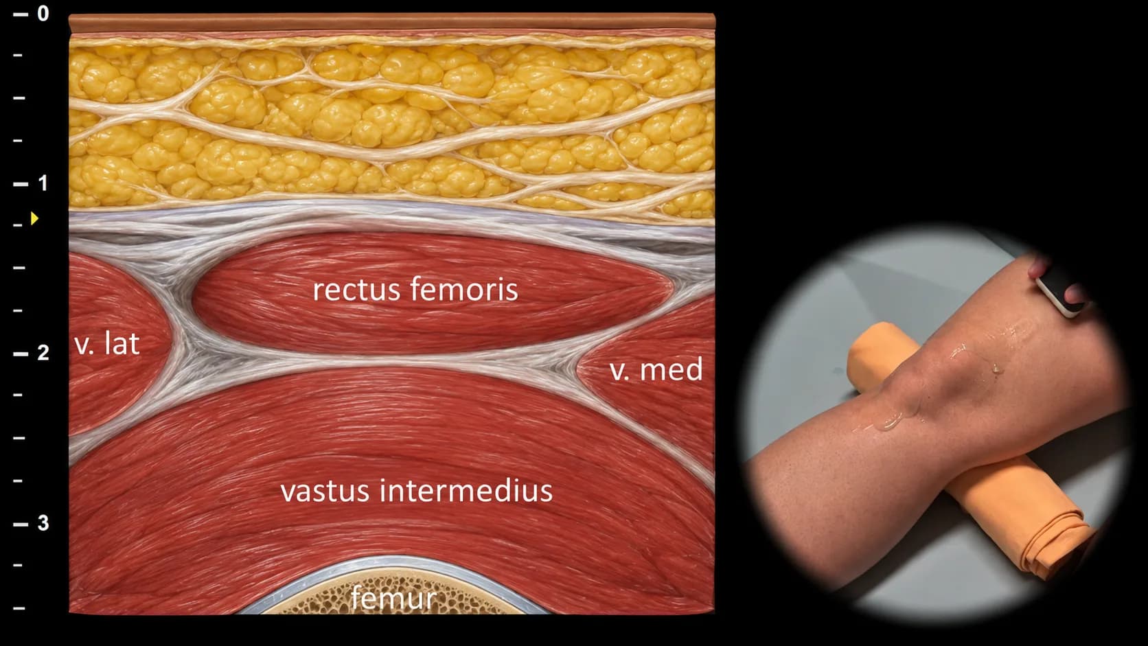
Figure 1. Ventral view, transverse plane, suprapatellar. v. lat: m. vastus lateralis, v. med: m. vastus medialis
Transverse ultrasound section of the anterior thigh at the suprapatellar level showing individual parts of the m. quadriceps femoris. Superficially in the midline, the m. rectus femoris is visible, with m. vastus lateralis and m. vastus medialis on its sides, and deeper the m. vastus intermedius, which lies against the anterior contour of the femur. The femur is displayed as a curved hyperechoic cortical line with dorsal acoustic shadowing. This projection is important for evaluating the anatomical arrangement of the quadriceps muscles, detecting muscle injuries, and providing orientation for subsequent tracking distally toward the quadriceps tendon.
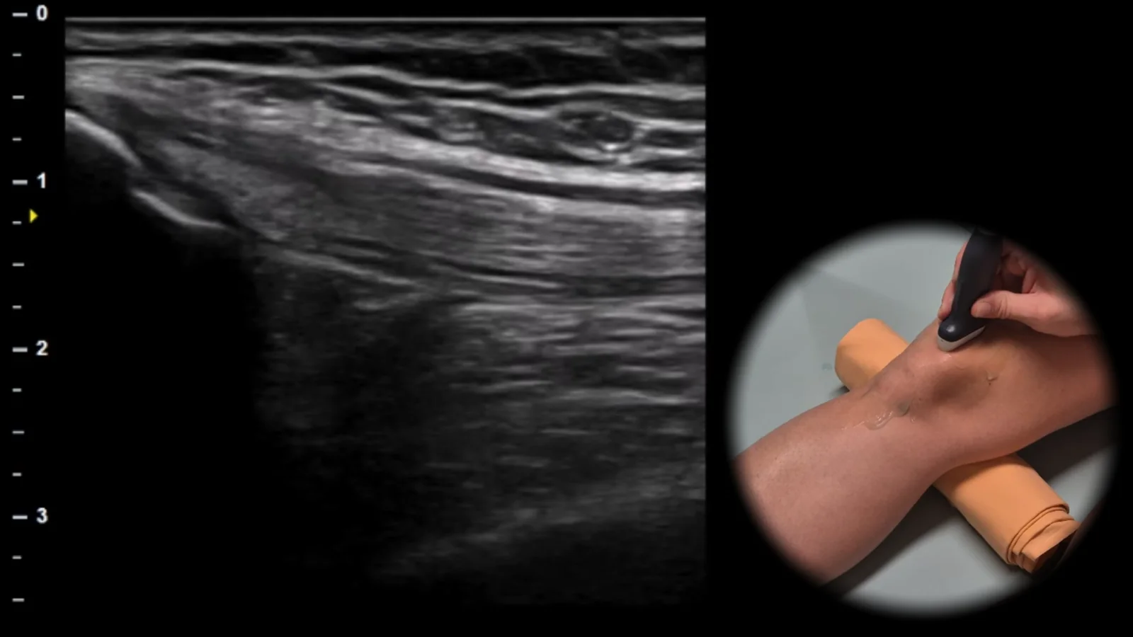
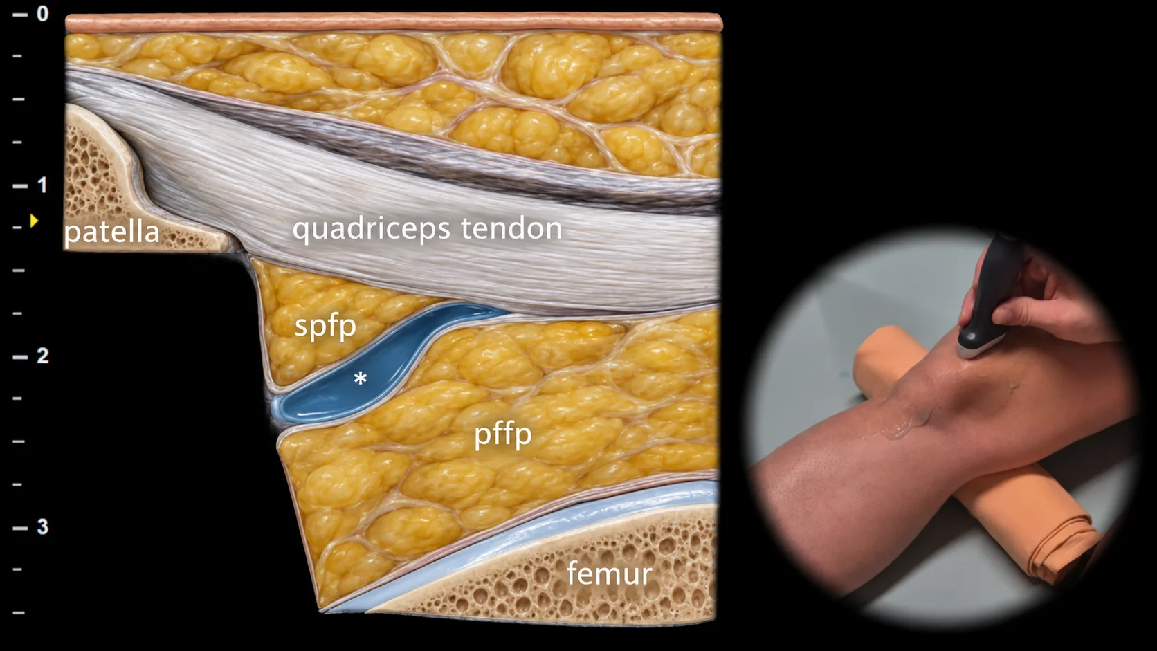
Figure 2. Ventral view, sagittal plane, suprapatellar. spfp: suprapatellar fat pad, pffp: prefemoral fat pad, *: suprapatellar recess
Longitudinal ultrasound section of the anterior part of the knee in the suprapatellar area. The quadriceps femoris tendon is shown in this projection as a broad hyperechoic fibrillar structure inserting on the superior pole of the patella. Deep beneath the tendon, the suprapatellar recess is visible as a potential space between the patella and femur, below which lies the prefemoral fat pad, while more superficially the suprapatellar fat pad is located, which together form important anatomical landmarks of this projection. This projection is crucial for evaluating the quadriceps tendon, suprapatellar recess, and signs of synovial pathology.
Clinical Note
In this area, fluid in the knee joint can be well detected, which accumulates in the suprapatellar recess and leads to its expansion in case of effusion.
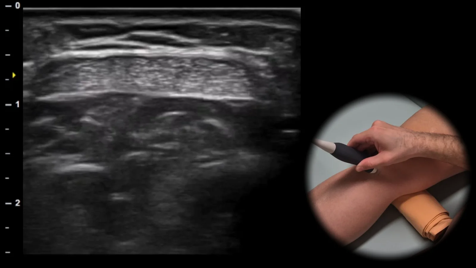
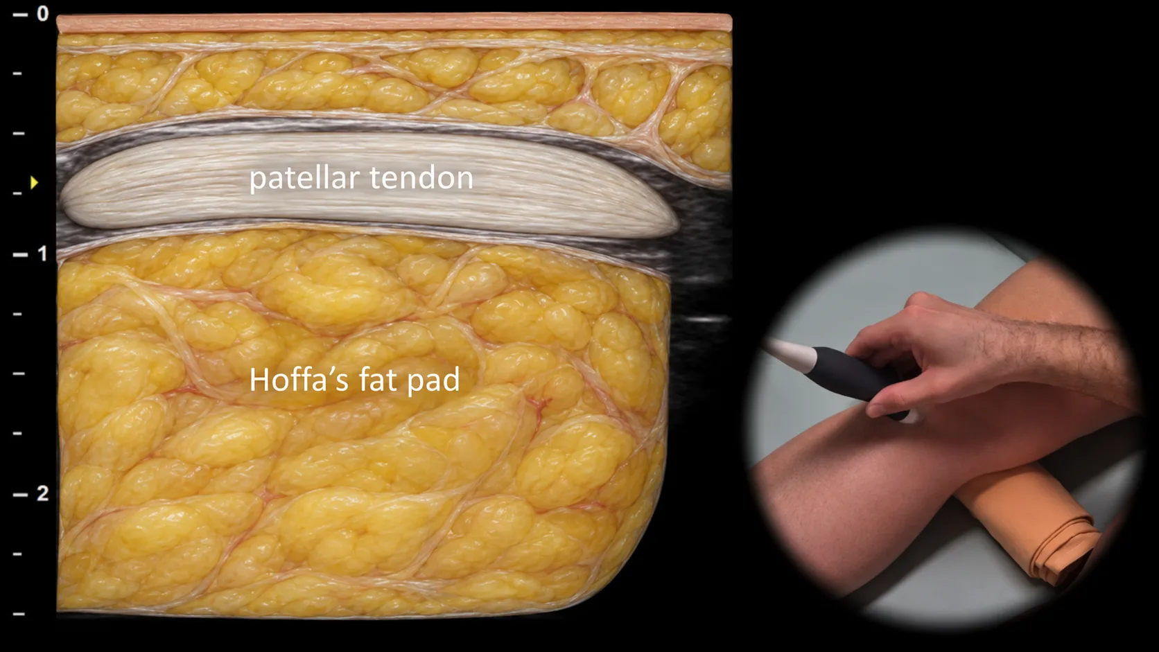
Figure 3. Ventral view, transverse plane, infrapatellar.
Transverse ultrasound section of the anterior part of the knee in the infrapatellar area. The patellar tendon is displayed in this projection as a hyperechoic fibrillar structure, beneath which Hoffa's fat pad is visible as an inhomogeneous soft tissue structure filling the anterior infrapatellar space. This projection is essential for assessment of the patellar tendon, detection of patellar tendinopathy, partial lesions, and changes in Hoffa's fat pad related to impingement or postoperative conditions.
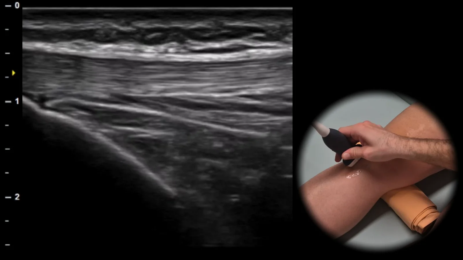
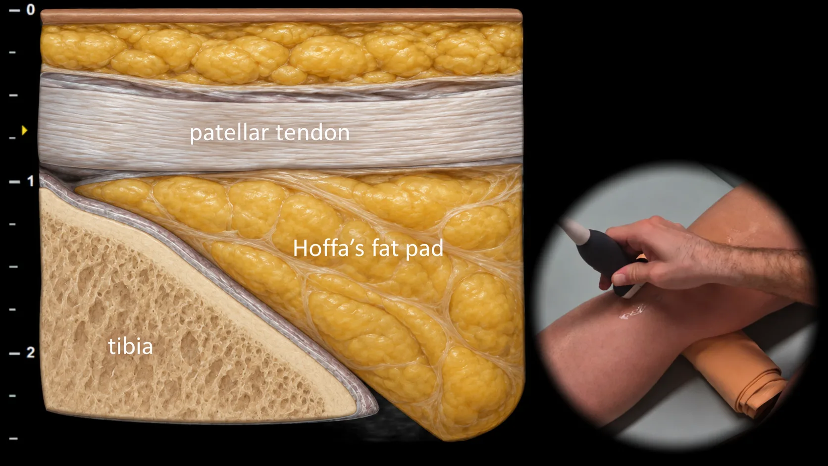
Figure 4. Ventral view, sagittal plane, infrapatellar.
Longitudinal ultrasound section of the anterior part of the knee in the infrapatellar region. The patellar tendon is displayed in this projection as a thick hyperechoic fibrillar band extending from the lower pole of the patella to its insertion on the anterior surface of the proximal tibia. Immediately below the tendon and in front of the tibia, Hoffa's fat pad is visible, which forms an important anatomical landmark of this region. This projection is essential for evaluating patellar tendinopathy, partial ruptures, and pathological changes in the area of the tibial insertion of the patellar tendon as well as Hoffa's fat pad.
Access the full Health Library
Full access to scanning protocols, anatomy, and clinical references. Cancel anytime.
- Every protocol and anatomy reference
- Original ultrasound illustrations and video demonstrations
- Sync across mobile and web