WRIST
Ultrasound allows immediate evaluation of periarticular soft tissues, both statically and dynamically. Following structures can be evaluated: tendons of flexors and extensor of the hand, radiocarpal joint, median and ulnar nerve, arteries.
Position of the patient:
Patient is seated in front of the examiner with a bed or a table in between. The position of the upper extremity varies from the scanning area (see figures below).
1. Anterior (palmar) view
A. Transversal plane
B. Longitudinal plane
C. Oblique plane
2. Medial (ulnar) view
A. Transversal plane (ECU)
3. Lateral (radial) view
A. Transversal plane
4. Posterior (dorsal) view
A. Transversal plane
B. Longitudinal plane
5. Finger view
A. Longitudinal plane
1. Anterior view
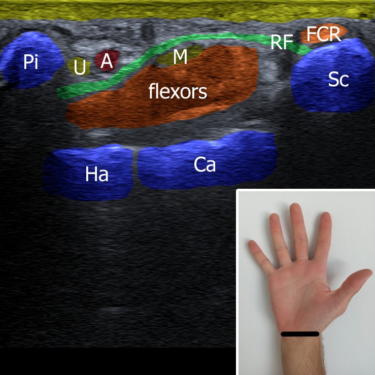
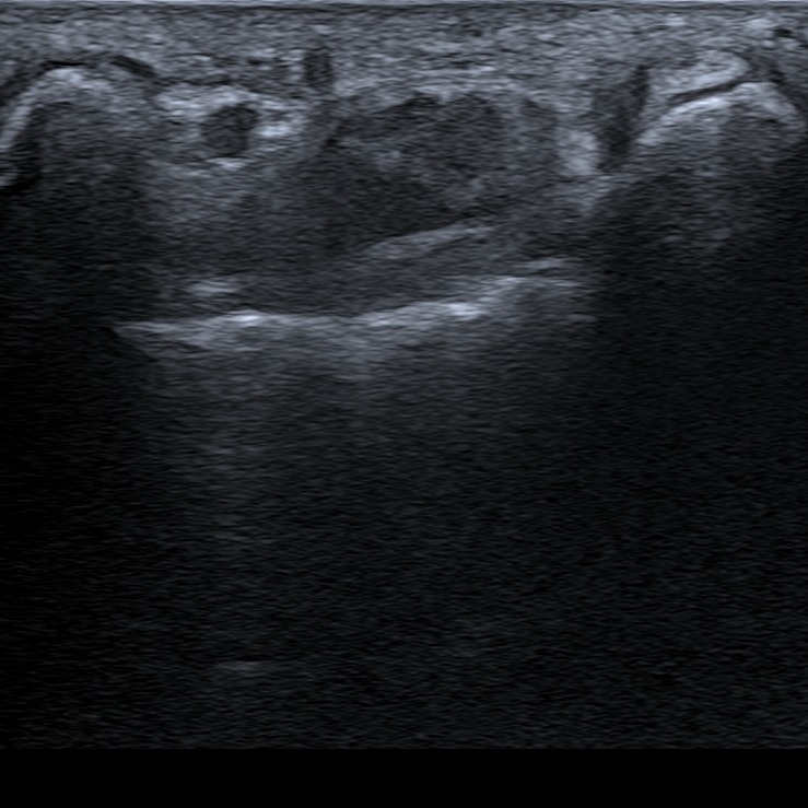
First of all carpal tunnel is located – the bony structures surrounding the carpal tunnel are pisiform, scaphoid, hamate and capitate bone. Tendons of the hand flexors and the median nerve are located in the carpal tunnel underneath the flexor retinaculum. At the ulnar side the Guyon’s canal can be seen with ulnar nerve and artery passing through.
Figure 1. U – ulnar nerve, M – median nerve, A – ulnar artery, Pi – os pisiforme, Ha – os hamatum, Ca – os capitatum, Sc – os scaphoideum, RF – retinaculum flexorum, FCR – flexor carpi ulnaris.
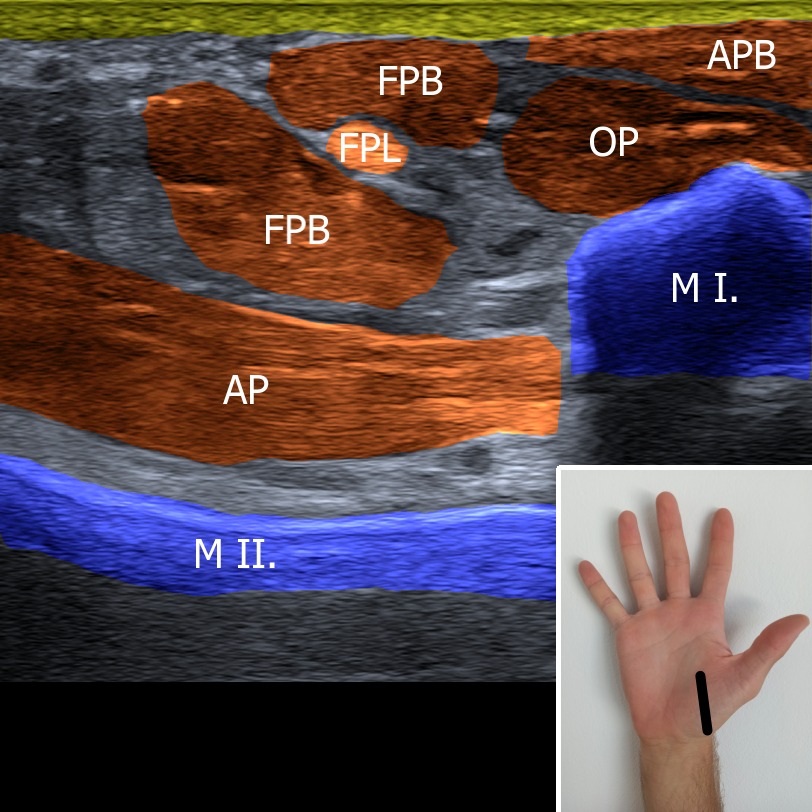
The probe is place at the thenar parallel with the long axis of the upper extremity. Tendon of the flexor pollicis longus can be seen as a “full moon sign” in it’s short axis between flexor pollicis brevis muscle.
Figure 2. M I. – first metacarp, M II. – second metacarp, AP – musculus adductor pollicis, FPB – musculus flexor pollicis brevis, FPL – musculus flexor pollicis longus , OP – musculus opponens pollicis, APB – musculus abductor pollicis brevis.
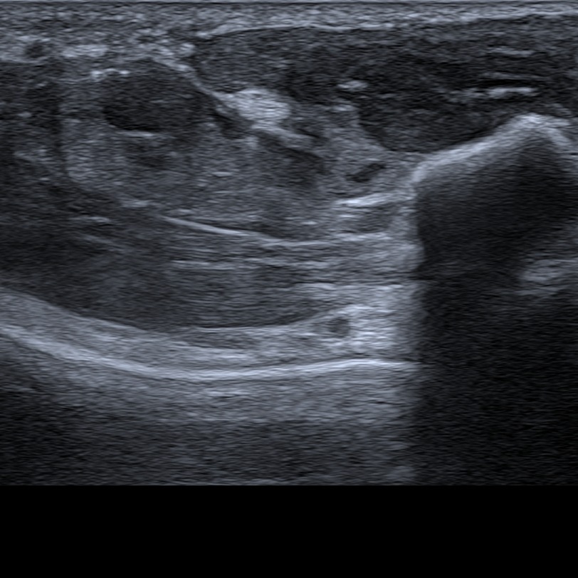
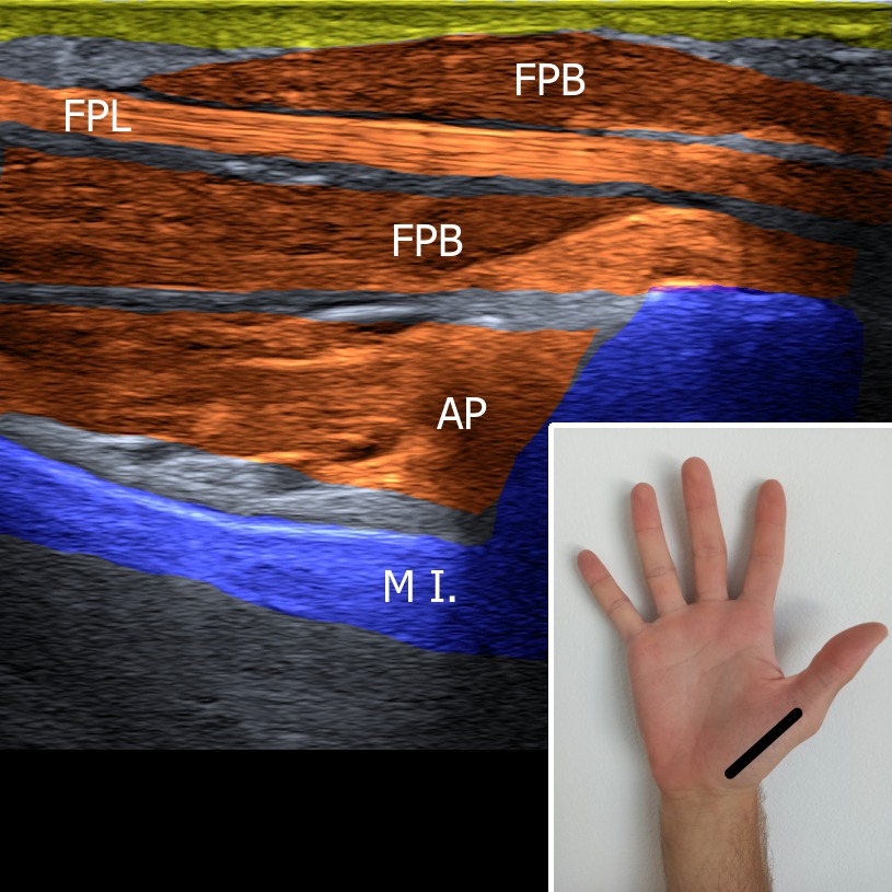
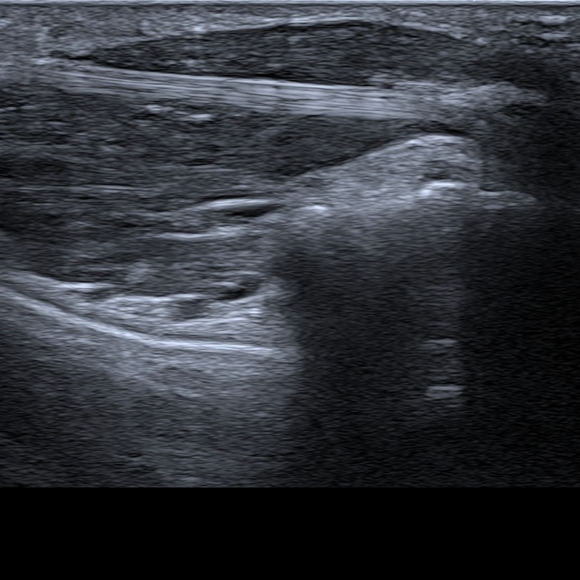
The tendon of the flexor pollicis longus can be also observed in the long axis. The tendon can be tracked till it’s distal insertion on the distal phalanx.
Figure 3. M I. – first metacarp, AP – musculus adductor pollicis, FPB – musculus flexor pollicis brevis, FPL – musculus flexor pollicis longus.
2. Medial (ulnar) view
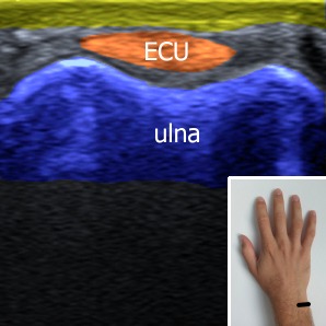
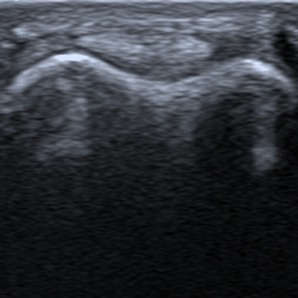
In this plane tendon of the extensor carpi ulnaris can be visualized as the 6th extensor compartment.
Figure 4. ECU – extensor carpi ulnaris.
3. Lateral (radial) view
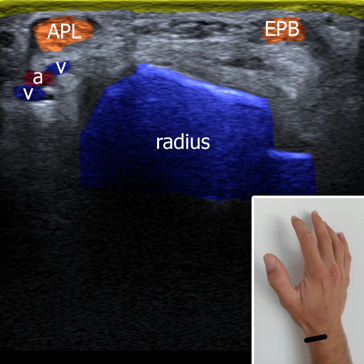
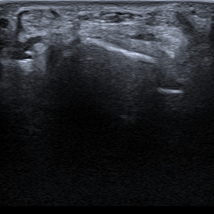
At the lateral (radial) side of the carpus 1st extensor comprment (abductor pollicis longus and extensor pollicis brevis) can be visualized. There can be also find tendon of extensor pollicis longus. At the bottom of the foveloa radialis pass radial artery and veins.
Figure 5. a – artery, v – vein, APB – musculus abductor pollicis longus, EPL – musculus extensor pollicis longus.
4. Posterior (dorsal) view
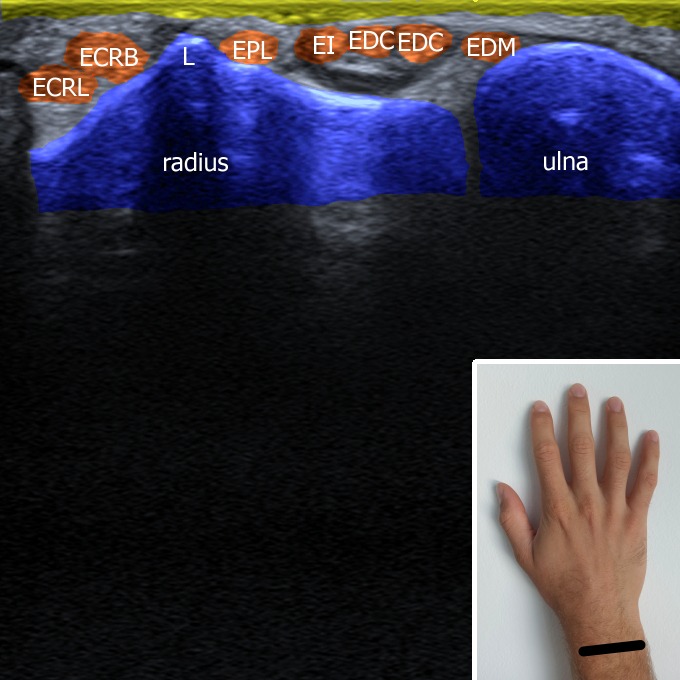
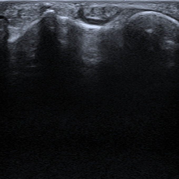
At the dorsal side the bony landmark to be looked for is Lister’s tubercle. This radial bone promimence separates the 2nd (extensor carpi radialis longus et brevis) and the 3rd (extensor pollicis longus) extensor compartment. 4th (extensor indicis et extensor digitorum communis) and 5th (extensor digiti minimi) are located at the ulnar side of the Lister’s tubercle.
Figure 6. L – Lister’s tubercle, ERCL – extensor carpi radialis longus, ECRB – extensor carpi radialis brevis, EPL – extensor pollicis longus, EI – extensor indicis, EDC – extensor digitorum communis, EDM – extensor digiti minimi.
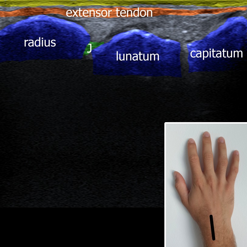
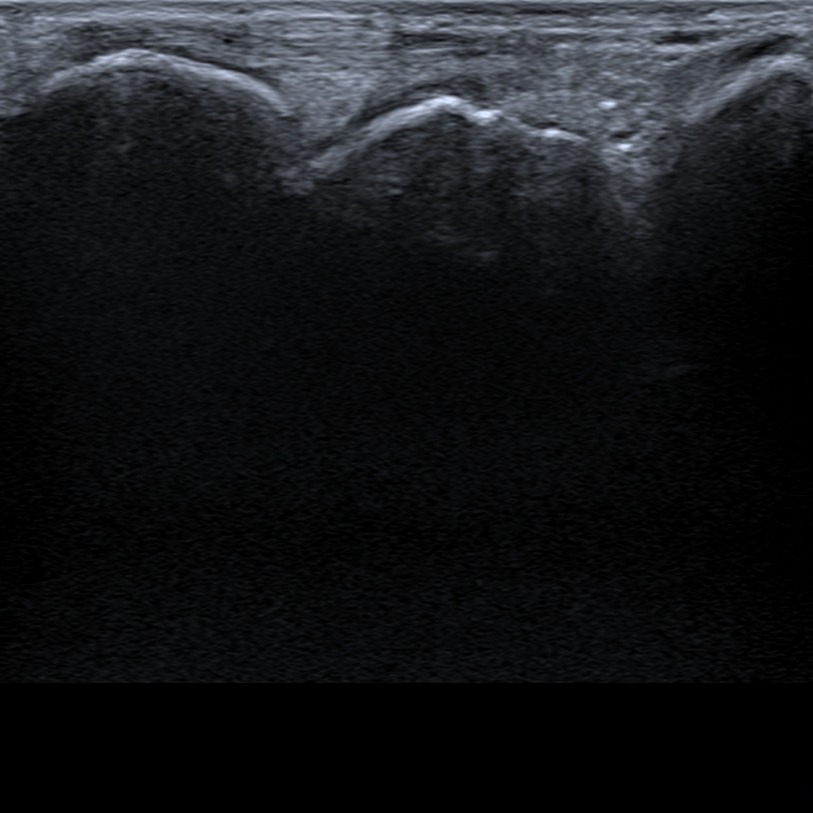
At this sonogram of radiocarpal joint is visualized a typical place where joint fluid can accumulate (articulation between radius and lunate).
Figure 7. J – radiocarpal joint.
5. Finger view
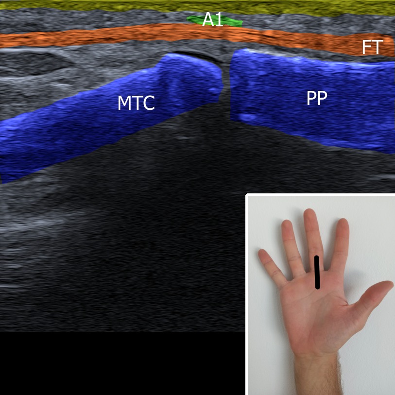
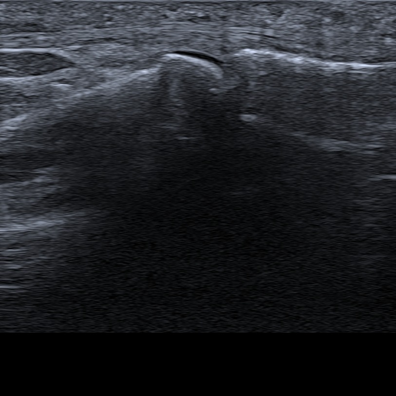
The probe is place in the long axis of the finger above metacarpophalangeal joint. In this plane flexor tendon of the finger and A1 pulley (hyperechoic structure above the joint) can be visualized.
Figure 8. A1 – A1 pulley, FT – flexor tendon, MTC – metacarpus, PP – proximal phalanx.
1. Anterior view
First of all carpal tunnel is located – the bony structures surrounding the carpal tunnel are pisiform, scaphoid, hamate and capitate bone. Tendons of the hand flexors and the median nerve are located in the carpal tunnel underneath the flexor retinaculum. At the ulnar side the Guyon’s canal can be seen with ulnar nerve and artery passing through.
Figure 1. U – ulnar nerve, M – median nerve, A – ulnar artery, Pi – os pisiforme, Ha – os hamatum, Ca – os capitatum, Sc – os scaphoideum, RF – retinaculum flexorum, FCR – flexor carpi ulnaris.


The probe is place at the thenar parallel with the long axis of the upper extremity. Tendon of the flexor pollicis longus can be seen as a “full moon sign” in it’s short axis between flexor pollicis brevis muscle.
Figure 2. M I. – first metacarp, M II. – second metacarp, AP – musculus adductor pollicis, FPB – musculus flexor pollicis brevis, FPL – musculus flexor pollicis longus , OP – musculus opponens pollicis, APB – musculus abductor pollicis brevis.


The tendon of the flexor pollicis longus can be also observed in the long axis. The tendon can be tracked till it’s distal insertion on the distal phalanx.
Figure 3. M I. – first metacarp, AP – musculus adductor pollicis, FPB – musculus flexor pollicis brevis, FPL – musculus flexor pollicis longus.


2. Medial (ulnar) view
In this plane tendon of the extensor carpi ulnaris can be visualized as the 6th extensor compartment.
Figure 4. ECU – extensor carpi ulnaris.


3. Lateral (radial) view
At the lateral (radial) side of the carpus 1st extensor comprment (abductor pollicis longus and extensor pollicis brevis) can be visualized. There can be also find tendon of extensor pollicis longus. At the bottom of the foveloa radialis pass radial artery and veins.
Figure 5. a – artery, v – vein, APB – musculus abductor pollicis longus, EPL – musculus extensor pollicis longus.
4. Posterior (dorsal) view
At the dorsal side the bony landmark to be looked for is Lister’s tubercle. This radial bone promimence separates the 2nd (extensor carpi radialis longus et brevis) and the 3rd (extensor pollicis longus) extensor compartment. 4th (extensor indicis et extensor digitorum communis) and 5th (extensor digiti minimi) are located at the ulnar side of the Lister’s tubercle.
Figure 6. L – Lister’s tubercle, ERCL – extensor carpi radialis longus, ECRB – extensor carpi radialis brevis, EPL – extensor pollicis longus, EI – extensor indicis, EDC – extensor digitorum communis, EDM – extensor digiti minimi.


At this sonogram of radiocarpal joint is visualized a typical place where joint fluid can accumulate (articulation between radius and lunate).
Figure 7. J – radiocarpal joint.


5. Finger view
The probe is place in the long axis of the finger above metacarpophalangeal joint. In this plane flexor tendon of the finger and A1 pulley (hyperechoic structure above the joint) can be visualized.
Figure 8. A1 – A1 pulley, FT – flexor tendon, MTC – metacarpus, PP – proximal phalanx.


1. Özçakar L, Kara M, Chang KV, Ulaşlı AM, Hung CY, Tekin L, Wu CH, Tok F, Hsiao MY, Akkaya N, Wang T, Çarli AB, Chen WS, De Muynck M. EURO-MUSCULUS/USPRM Basic Scanning Protocols for wrist and hand. Eur J Phys Rehabil Med. 2015 Aug;51(4):479-84. Epub 2015 Jul 9. PMID: 26158917.
