Metaphoric Signs in msk Sonography – Physiology
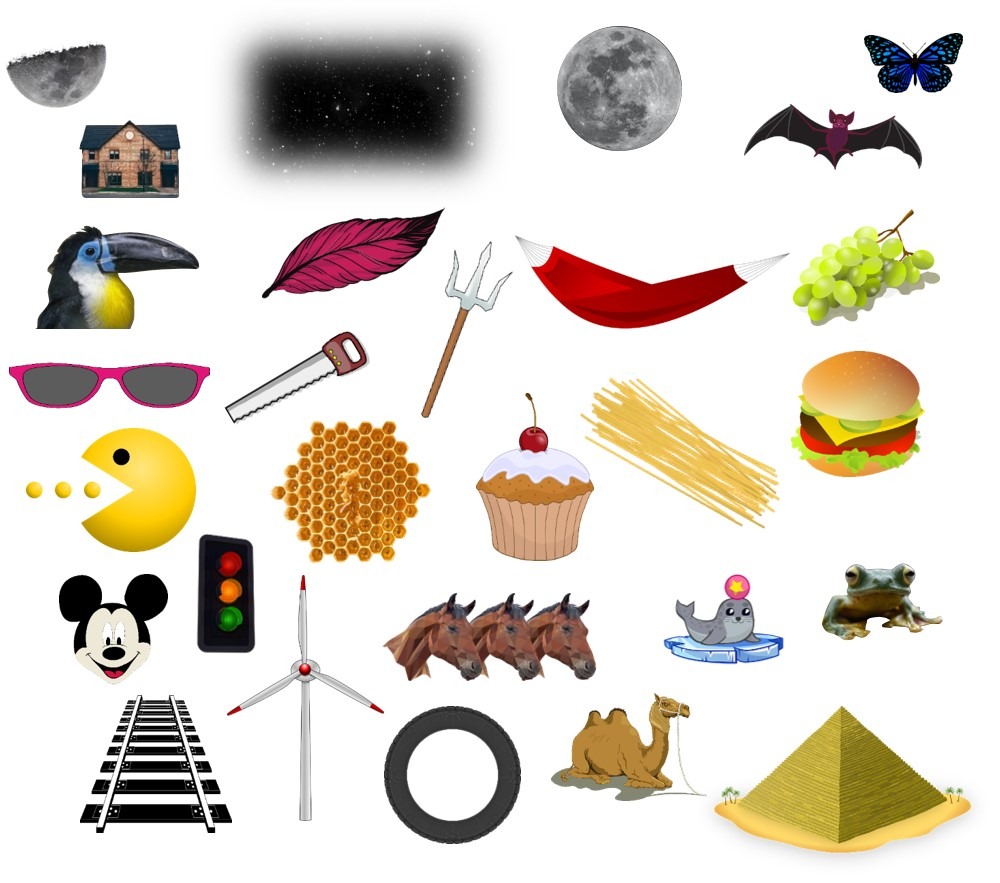
This post is dedicated to article called Mnemonics and Metaphorical Videos for Teaching/Learning Musculoskeletal Sonoanatomy. The article was published in American Journal of Physical Medicine and Rehabilitation in December 2022.
In this article, the authors describe a new approach to teaching musculoskeletal ultrasonography. The authors have created a collection of images, drawings, and multimedia videos that use mnemonic aids, pictures, and characteristic sounds to help novice sonographers understand and remember the topographic anatomy of the musculoskeletal system. This approach, called “entertainment education,” combines text and video to provide a clear and engaging explanation of procedures in musculoskeletal medicine.
I. General
1. Muscle – Starry sky
2. Muscle – Feather
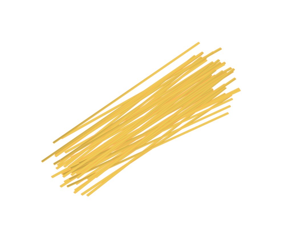
3. Tendon – Spaghetti
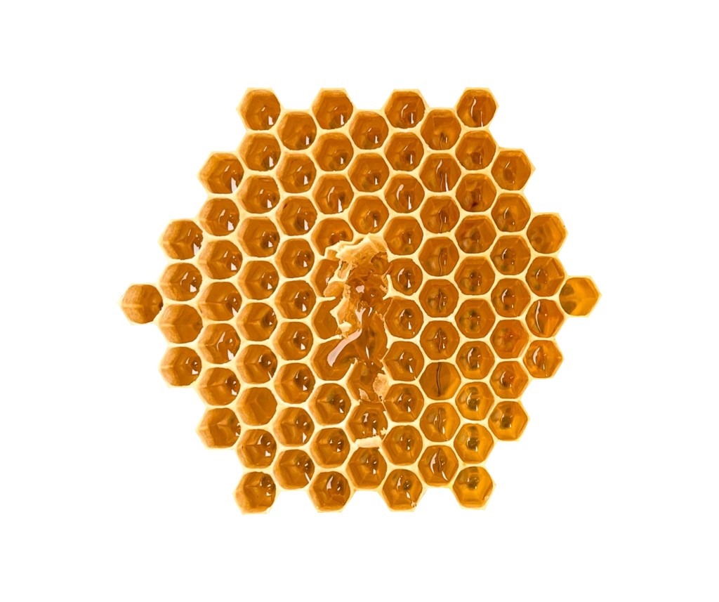
4. Nerve – Honeycomb
5. Vessels – Mickey Mouse
II. Specific
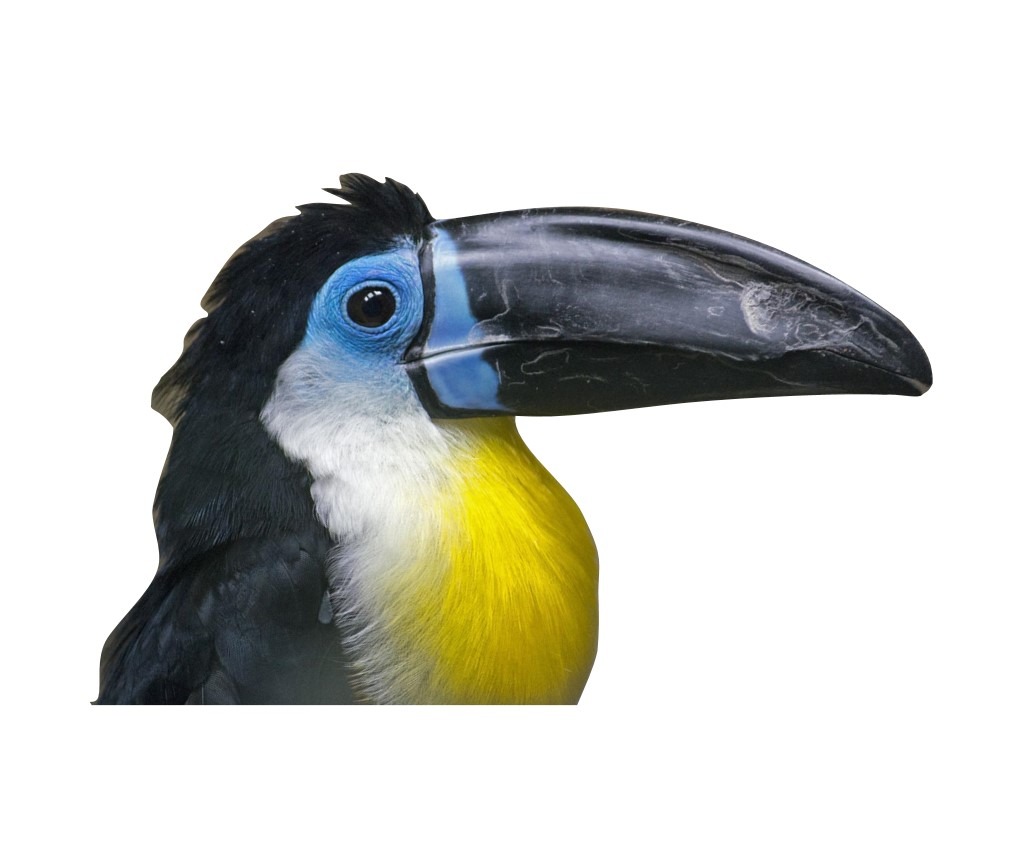
1. Rotator cuff (longitudinal) – Bird beak
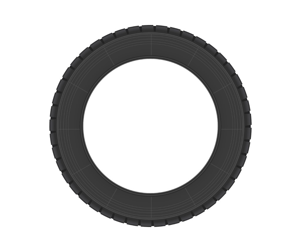
2. Rotator cuff (transversal) – Tire
3. Pronator teres + brachialis + median nerve – Hamburger
4. Flexor carpi ulnaris + ulna – Moon over a house
5. Flexor pollicis longus tendon – Full moon
6. Greater trochanter – Pyramid
7. Greater trochanter – Matterhorn
8. Sciatic nerve + tendons – Windmill
9. Sciatic nerve + tendons – Mercedes
10. Short head of biceps femoris – Railway
11. Semimembranosus + semitendinosus – Cherry on the cake
12. Gastrocnemius – Sunglasses
13. Tibialis posterior – Seal with a ball
14. Calcaneofibular ligament – Hammock
15. Plantar muscles and tendons – Pac-Man
16. Nerve roots C5, C6 nd C7 – Traffic lights
17. Brachial plexus – Grapes
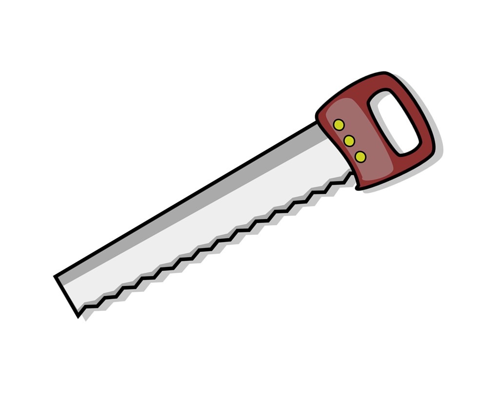
18. Cervical spine facet joints – Saw teeth
19. Lubmar spine – transverse processes – Trident
20. Lumbar spine facet joints – Camel humps
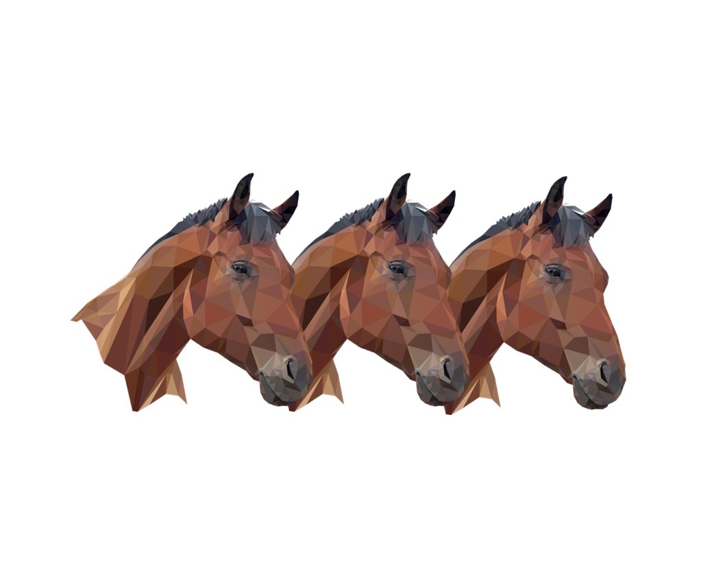
21. Lumbar spine laminae – Horse heads
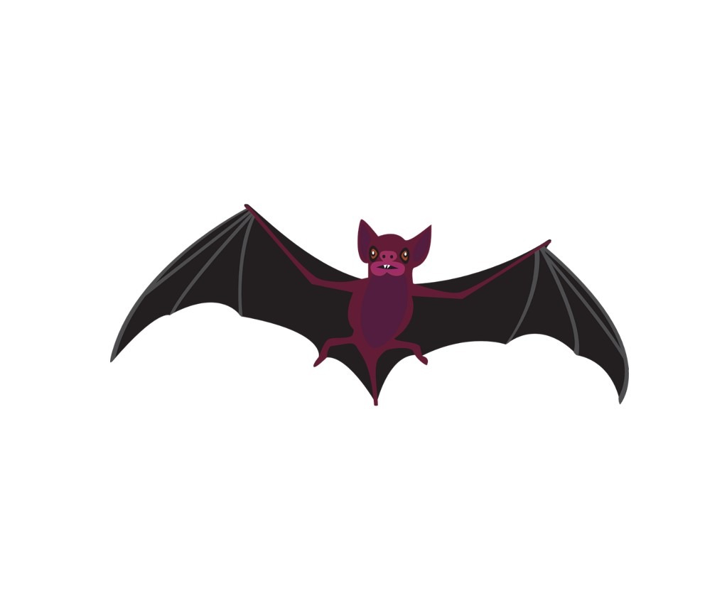
22. Lumbar vertebra – Bat
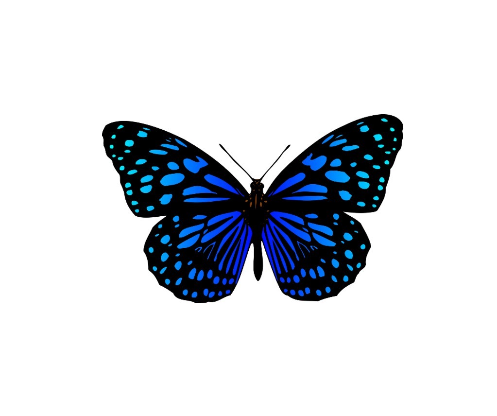
23. Lumbar paraspinal muscles – Butterfly
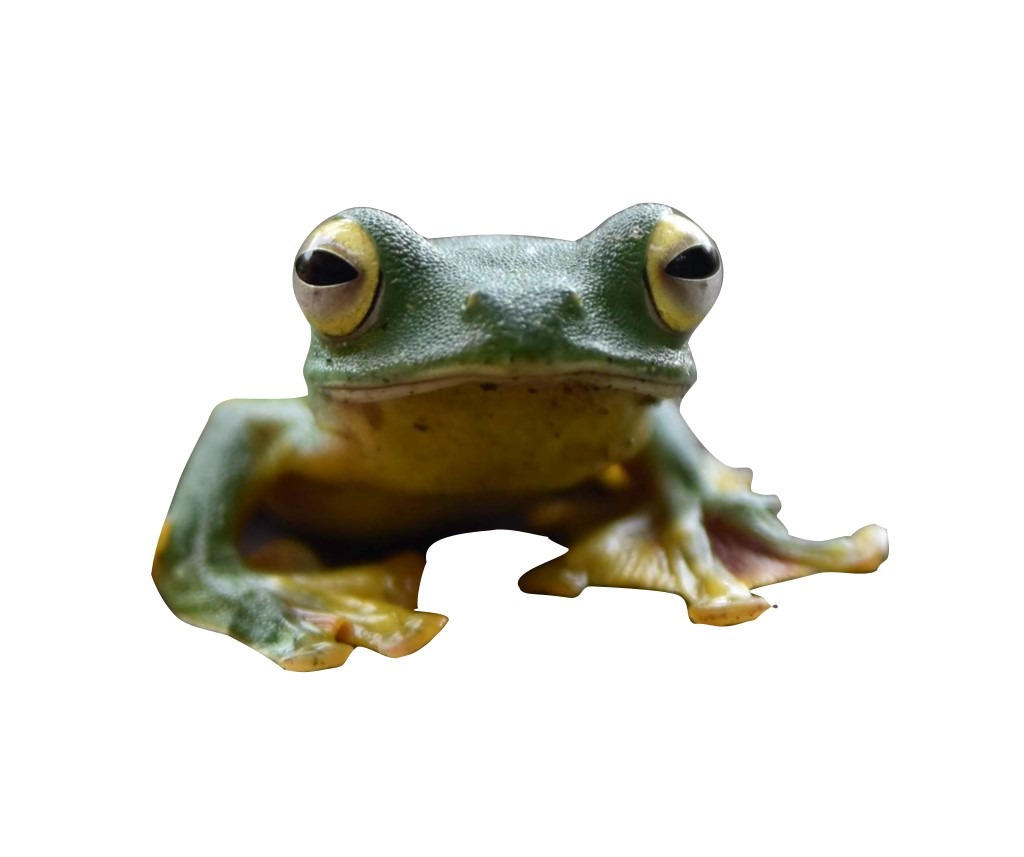
24. Cornua sacralia – Frog eyes
General
Muscle – Starry sky – video link
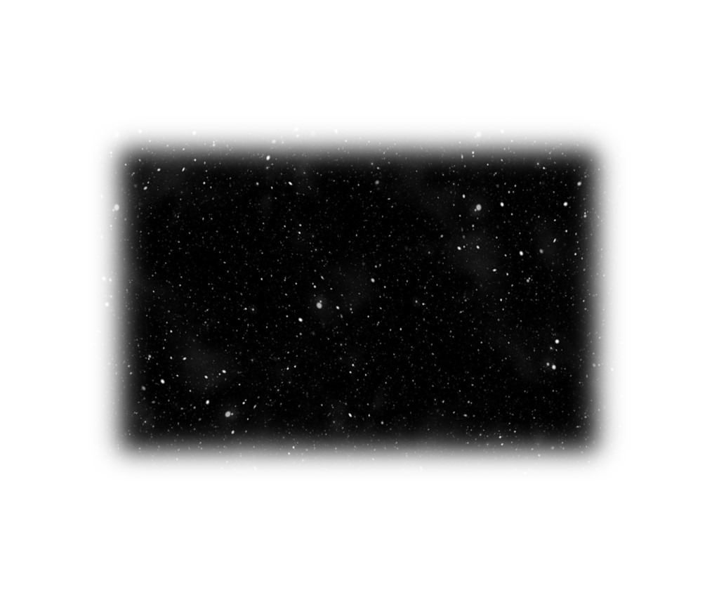
Muscles have a specific internal structure made up of hypoechoic fascicles (dark areas) and hyperechoic perimysium (bright areas). When examining muscles using transverse scans, these two compartments can be observed alternating with one another. The hypoechoic fascicles resemble the dark sky, while the hyperechoic perimysium resembles stars. The appearance of this “relaxing starry sky” pattern is often used as a way to identify muscle tissue during diagnostic examinations. The presence or absence of this pattern can help determine if a muscle is normal or abnormal.
Muscle – Feather – video link
Muscle fascicles are long, thin fibers that run either parallel to the length of the muscle (in fusiform muscles) or at an angle to the aponeurosis (in pennate muscles). When examining a bipennate muscle using a longitudinal scan, the hypoechoic muscular tissue can be seen to contain branches of hyperechoic fibroadipose tissue. This pattern is similar to the structure of a feather, with a central beam and multiple peripheral branches. Recognizing this pattern can be helpful in accurately measuring the architecture of the muscle.

Tendon – Spaghetti – video link
Nerve – Honeycomb – video link
Vessels – Mickey Mouse – video link

Specific
Rotator cuff (longitudinal) – Bird beak – video link
The supraspinatus tendon is a long, thin structure that runs from the top of the shoulder blade, under the acromioclavicular joint, and attaches to the upper arm bone. When examining the supraspinatus tendon using a longitudinal scan with the shoulder in a neutral position, it may resemble the beak of a bird, with the acromion corresponding to the head of the bird. This strong “beak” helps to prevent the humeral head from sliding upward and becoming dislocated from the acromion (a condition known as cranial subluxation). The appearance of a normal supraspinatus tendon, as seen through this “beak” pattern, may indicate the presence of a healthy tendon in the critical zone.
Rotator cuff (transversal) – Tire – video link
Pronator teres + brachialis + median nerve – Hamburger – video link

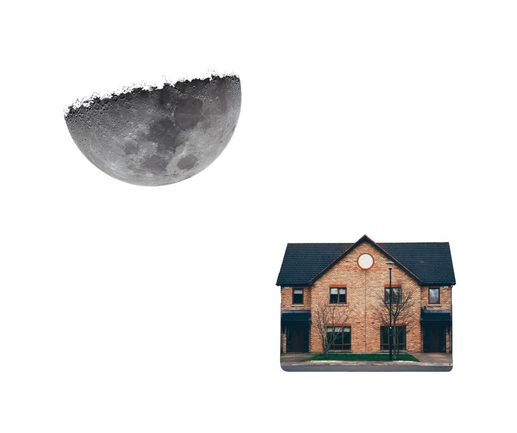
Flexor carpi ulnaris + ulna – Moon over a house – video link
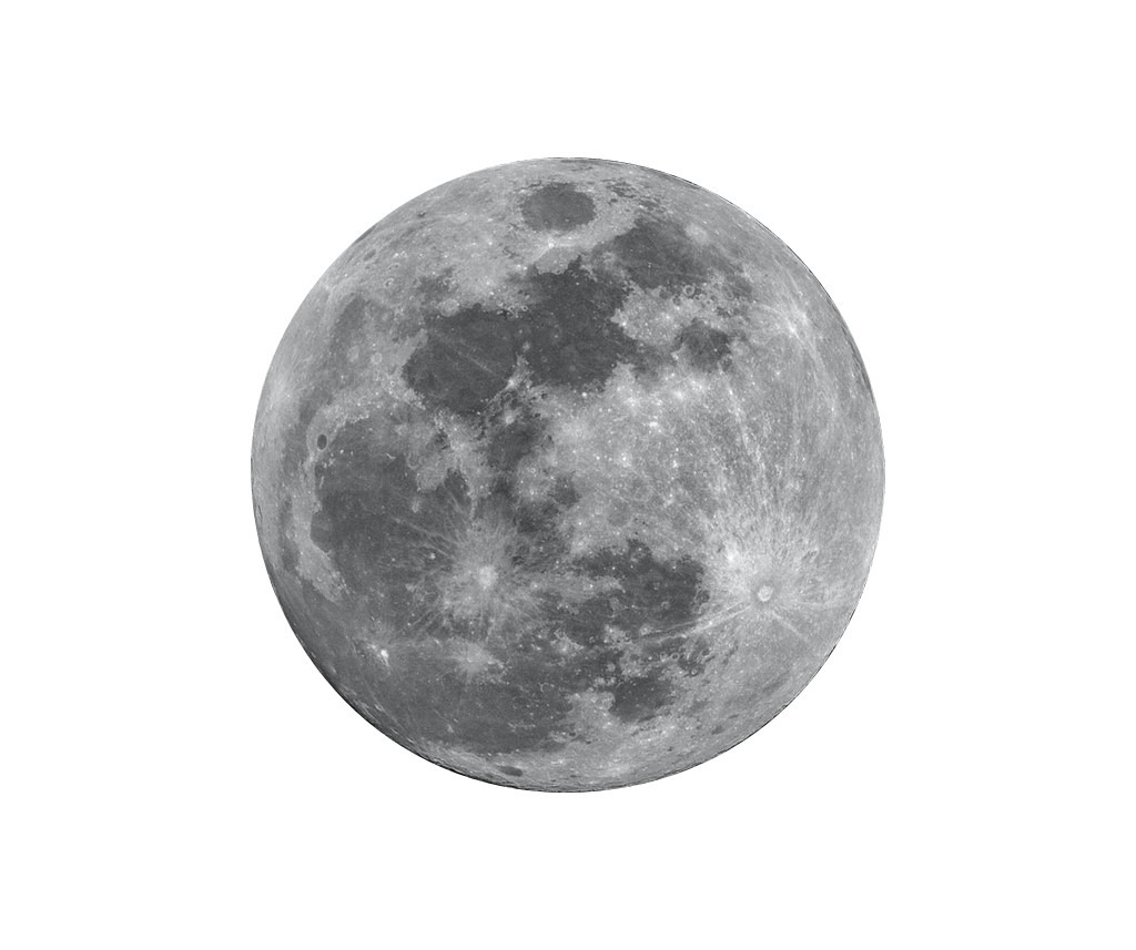
Flexor pollicis longus tendon – Full moon – video link
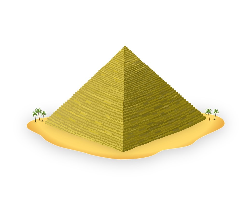
Greater trochanter – Pyramid – video link
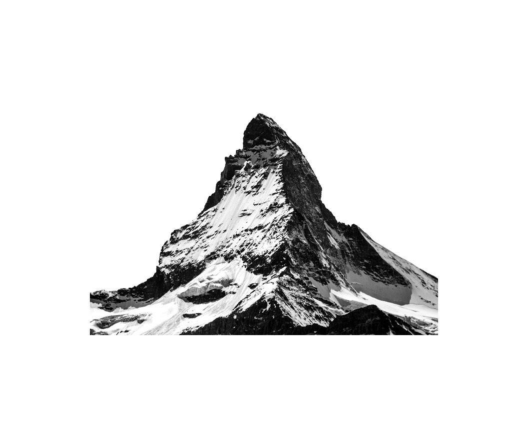
Greater trochanter – Matterhorn
The greater trochanter is a bony prominence on the outside of the hip that can be important for evaluating the lateral hip region. When examining this area using a transverse scan, the greater trochanter may resemble a triangular, hyperechoic shape similar to Mtterhorn. Recognizing this shape can be helpful for identifying the different facets of the greater trochanter and for distinguishing it from the rounder shape of the femoral shaft.
Sciatic nerve + tendons – Windmill – video link
Sciatic nerve + tendons – Mercedes
When examining the top third of the back of the thigh using a transverse scan, it is possible to see a “windmill” pattern formed by the conjoined tendon of the semitendinosus and biceps femoris muscles, the sciatic nerve, and the adductor magnus muscle. These anatomical structures, which metaphorically resemble the blades of a windmill, play a crucial role in providing the “energy” for lower limb movements. Recognizing this pattern can help physicians accurately locate the sciatic nerve and avoid injury. This pattern has also been referred to as the “Mercedes-Benz sign” in the literature, but in the future it may be more commonly referred to as the “windmill” pattern.

Short head of biceps femoris – Railway – video link
Semimembranosus + semitendinosus – Cherry on the cake – video link

Gastrocnemius – Sunglasses – video link
Tibialis posterior – Seal with a ball – video link
The tibialis posterior muscle is a deep muscle located on the back and inner side of the leg. It is covered by the flexor hallucis longus and flexor digitorum longus muscles. When examining this area using a transverse scan, it is possible to see the tibialis posterior muscle, the posterior tibial artery, and the tibial nerve, which together may resemble a sweet seal with a ball. Identifying these structures is important in order to accurately target the right muscle and avoid the nerve and artery when administering botulinum toxin injections or other interventions in this region.

Calcaneofibular ligament – Hammock – video link
The calcaneofibular ligament is a band of tissue that is part of the lateral ligament complex in the ankle. It runs from the lateral malleolus (the bony bump on the outside of the ankle) to the tubercle on the outer side of the heel bone (calcaneus). To visualize the calcaneofibular ligament, it may be helpful to perform a longitudinal (oblique) scan. The ligament may appear like a comfortable hammock on which the fibularis brevis and longus tendons (two muscles in the lower leg) swing.
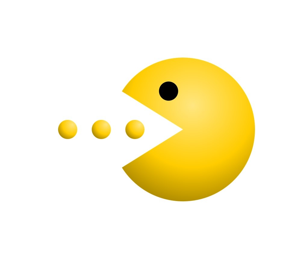
Plantar muscles and tendons – Pac-Man – video link
The plantar intrinsic muscles of the foot are located near the tendons of the extrinsic muscles of the foot. When examining the sole of the foot using a transverse scan, it is possible to see the plantar intrinsic muscles resembling a hungry Pac-Man, with the flexor digitorum longus and flexor hallucis longus tendons nearby. If these tendons are traced proximally (toward the body), their point of crossing, known as the “knot of Henry,” can be easily visualized. Identifying this knot can be important during the examination of plantar intersection syndrome, a condition that affects the tendons and muscles in the foot.
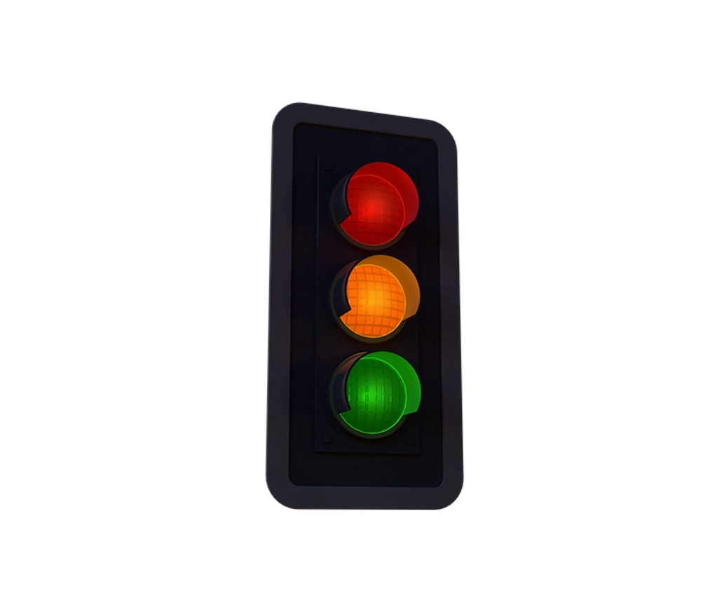
Nerve roots C5, C6 nd C7 – Traffic lights – video link
After leaving the neural foramina (small openings in the bones of the spine), the cervical nerve roots run between the scalene muscles in the neck. When examining the side of the neck from top to bottom using a transverse scan, it is possible to see the C5, C6, and C7 nerve roots positioned between the anterior and middle scalene muscles, resembling a set of traffic lights. Recognizing the location of these nerve roots can be crucial for a variety of neck interventions.
Brachial plexus – Grapes – video link
In the suprascapular region (the area above the shoulder blade), the cervical nerve roots come together to form trunks, which are then divided into upper and lower divisions. When examining the side of the neck using a transverse scan, it is possible to see the brachial plexus (a network of nerves that supplies the upper limb) running between the anterior and middle scalene muscles. This pattern may resemble a bunch of grapes. Recognizing the location of the brachial plexus is important for various neck procedures.

Cervical spine facet joints – Saw teeth – video link
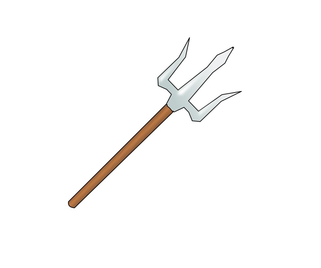
Lubmar spine – transverse processes – Trident – video link
When examining the lower back using a longitudinal paramedian scan, it is possible to see the three transverse processes (small bony projections from the sides of the vertebrae) that may create sharp shadowings. Recognizing this pattern, which may resemble a trident, can help ensure that the imaging provides a far lateral view of the spine. This can be useful for targeting the lumbar roots (the nerves that emerge from the lower back) during interventional procedures.
Lumbar spine facet joints – Camel humps – video link
Each lumbar vertebra (a bone in the lower back) consists of a body, arch (made up of two laminae and two pedicles), and two transverse processes and one spinous process (bony projections from the back of the vertebra). When examining the lower back using longitudinal paramedian scans, it is possible to see the facet joints and the laminae, which may resemble camel humps and horse heads, respectively. These structures are often targeted during relevant interventions.

Lumbar spine laminae – Horse heads – video link
Each lumbar vertebra (a bone in the lower back) consists of a body, arch (made up of two laminae and two pedicles), and two transverse processes and one spinous process (bony projections from the back of the vertebra). When examining the lower back using longitudinal paramedian scans, it is possible to see the facet joints and the laminae, which may resemble camel humps and horse heads, respectively. These structures are often targeted during relevant interventions.
Lumbar vertebra – Bat – video link
When examining the lumbar vertebrae using a transverse scan, it is possible to see the deep bony lining and the erector spinae muscles. These structures may resemble a bat and a butterfly, respectively. The bony structures serve as important landmarks during relevant interventions, and imaging the erector spinae muscles may also be useful during exercise therapy when using “sono-feedback” (a technique that involves using ultrasound images to provide visual feedback to guide muscle training).
Lumbar paraspinal muscles – Butterfly – video link
When examining the lumbar vertebrae using a transverse scan, it is possible to see the deep bony lining and the erector spinae muscles. These structures may resemble a bat and a butterfly, respectively. The bony structures serve as important landmarks during relevant interventions, and imaging the erector spinae muscles may also be useful during exercise therapy when using “sono-feedback” (a technique that involves using ultrasound images to provide visual feedback to guide muscle training).
Cornua sacralia – Frog eyes – video link
The sacrum is a bone formed by the fusion of the sacral vertebrae and is the continuation of the vertebral canal. The 5th sacral laminae do not fuse, resulting in a bony defect known as the sacral hiatus. The lateral walls of the sacral hiatus are made up of the tubercles of the inferior articular processes of the 5th sacral vertebrae, known as the sacral cornua. When examining this area using a transverse scan, it is possible to see the sacral cornua, which may resemble frog eyes. Identifying the hyperechoic band between these “eyes,” which is known as the sacrococcygeal ligament, can be important when planning ultrasound-guided procedures in this region.
1. Jačisko J, Mezian K, Güvener O, Ricci V, Kobesová A, Özçakar L. Mnemonics and Metaphorical Videos for Teaching/Learning Musculoskeletal Sonoanatomy. Am J Phys Med Rehabil. 2022 Dec 1;101(12):e189-e193. doi: 10.1097/PHM.0000000000002084. Epub 2022 Aug 9. PMID: 35944076.
