 MSK Academy
2. Lateral view
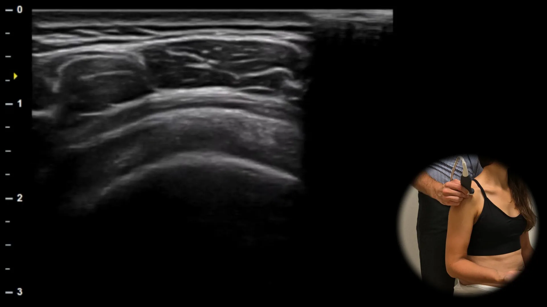
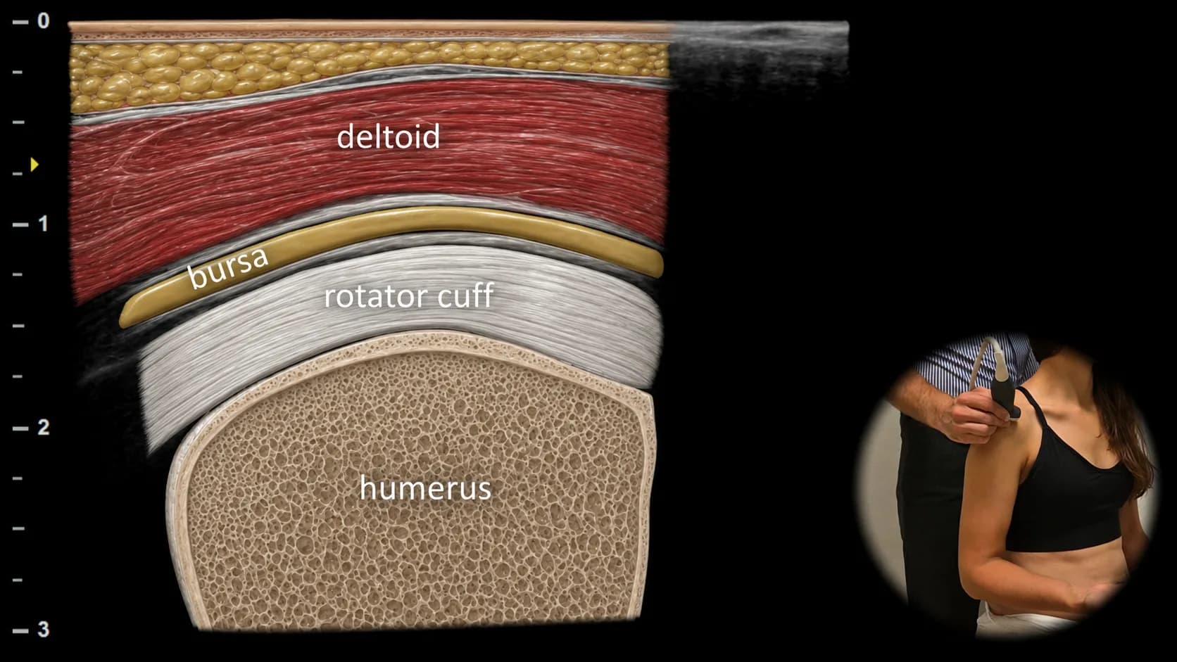
Figure 1. Lateral view, transverse plane.
Transverse ultrasound section of the lateral part of the shoulder showing the rotator cuff tendon in short axis, typically referred to as the "tire sign". In this projection, the integrity of the tendon can be assessed with gentle probe compression, where a healthy tendon maintains its firmness, resists pressure and has a rounded shape, while in case of rupture or significant damage it tends to be soft, easily compressible and may resemble a "flat tire". This projection is important for assessing continuity of the rotator cuff tendon and for detecting focal pathological changes, such as tears or calcifications, which may be visible only in part of its course.
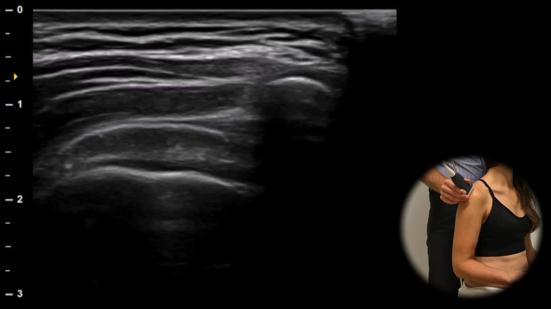
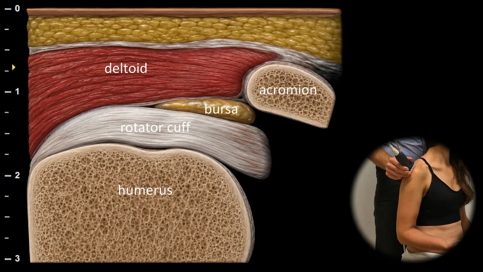
Figure 2. Lateral view, frontal plane.
Longitudinal ultrasound section of the lateral part of the shoulder showing the acromion and greater tubercle of the humerus as main bony landmarks. Between them, the supraspinatus tendon is visible in long axis with its typical "bird's beak" shape, and for complete evaluation of its approximately 4 cm wide structure, it is necessary to move the probe in an anteroposterior direction. Above the tendon lies the subacromial-subdeltoid bursa, which becomes more apparent when enlarged or inflamed. This projection is essential for evaluating tendinopathy, partial and complete ruptures of the supraspinatus tendon, and signs of subacromial impingement.