 MSK Academy
1. Ventral view
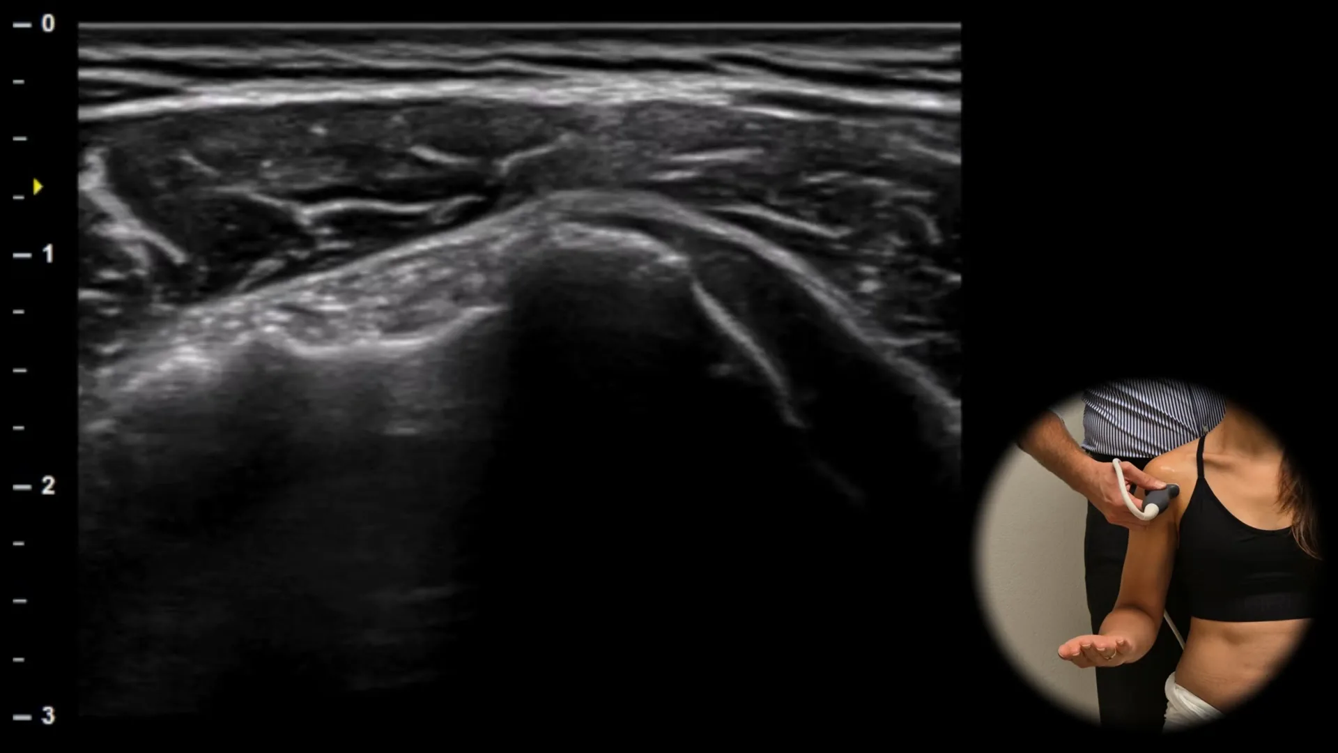
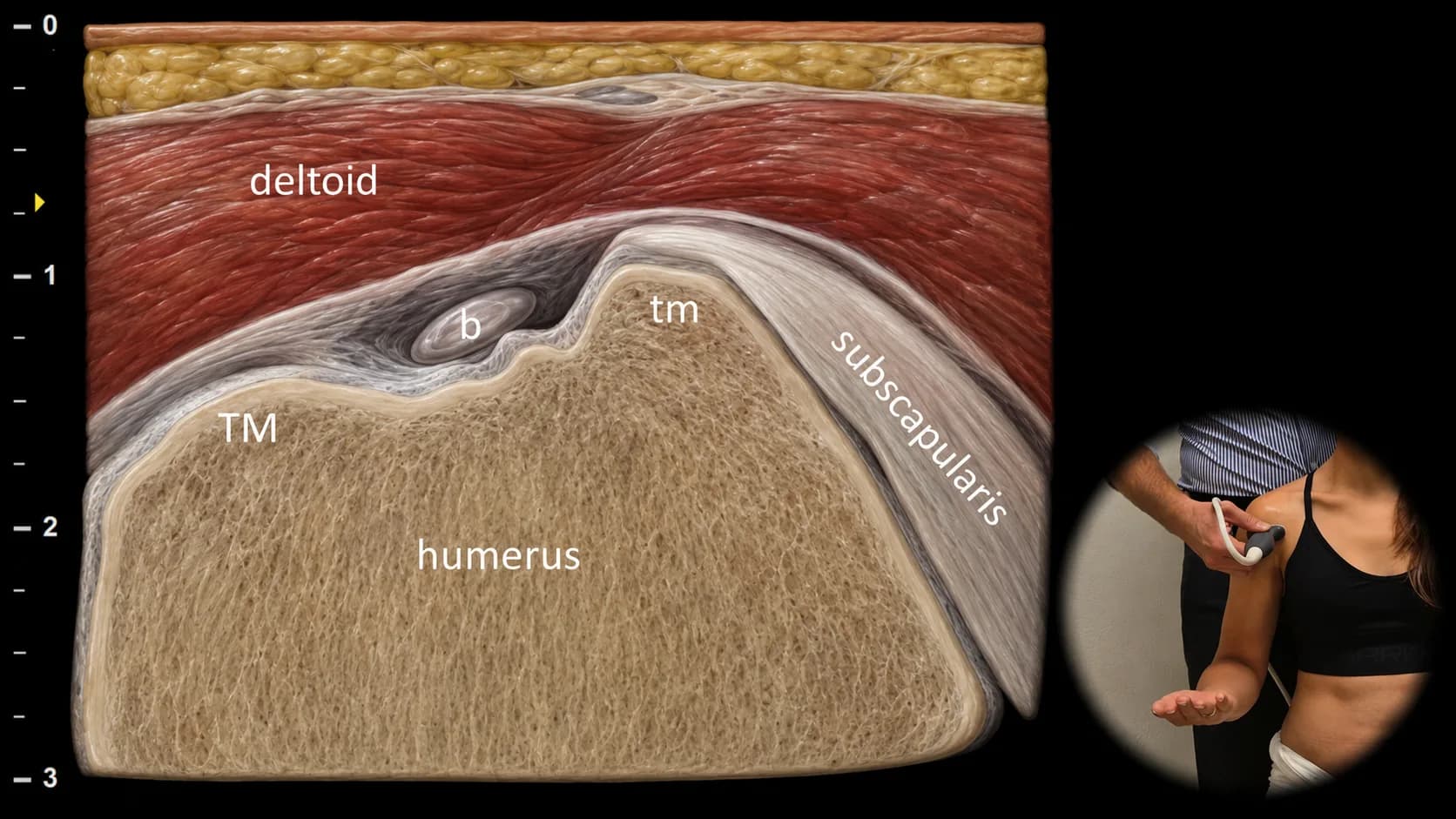
Figure 1. Ventral view, transverse plane. b: tendon of the long head of biceps brachii, TM: tuberculum majus, tm: tuberculum minus
Transverse ultrasound section of the anterior part of the shoulder joint. The proximal humerus is displayed with the greater (TM) and lesser (tm) tubercle, between which the tendon of the long head of biceps brachii (b) is located in the intertubercular sulcus. In the surrounding area, the tendon of subscapularis inserting on the lesser tubercle of the humerus and the superficially located deltoid muscle are also visible, whereby this projection provides an overview of the main anatomical landmarks of the anterior part of the shoulder and enables targeted evaluation of tendons, soft tissues, and any pathological changes.
Clinical Note
The area around the long head of biceps tendon represents a typical site of fluid accumulation, because the tendon sheath communicates with the joint cavity; the presence of fluid in this location can therefore be a sign of effusion in the shoulder joint and requires careful assessment of its extent and character.

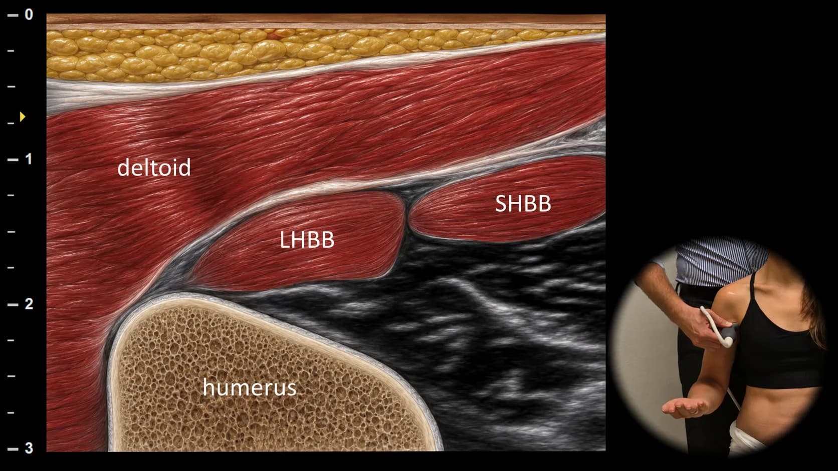
Figure 2. Ventral view, transverse plane. LHBB: long head of biceps brachii m., SHBB: short head of biceps brachii m.
Transverse ultrasound section of the anterior part of the arm with distal movement of the probe along the biceps brachii m. In this projection, both heads of the muscle are clearly visible, the long head (LHBB) and short head (SHBB), which form the muscle belly in the anterior region of the arm. This projection is suitable for assessing the structure and symmetry of the muscle belly and for detecting pathological changes such as partial or complete ruptures, hematomas, or signs of muscle atrophy.
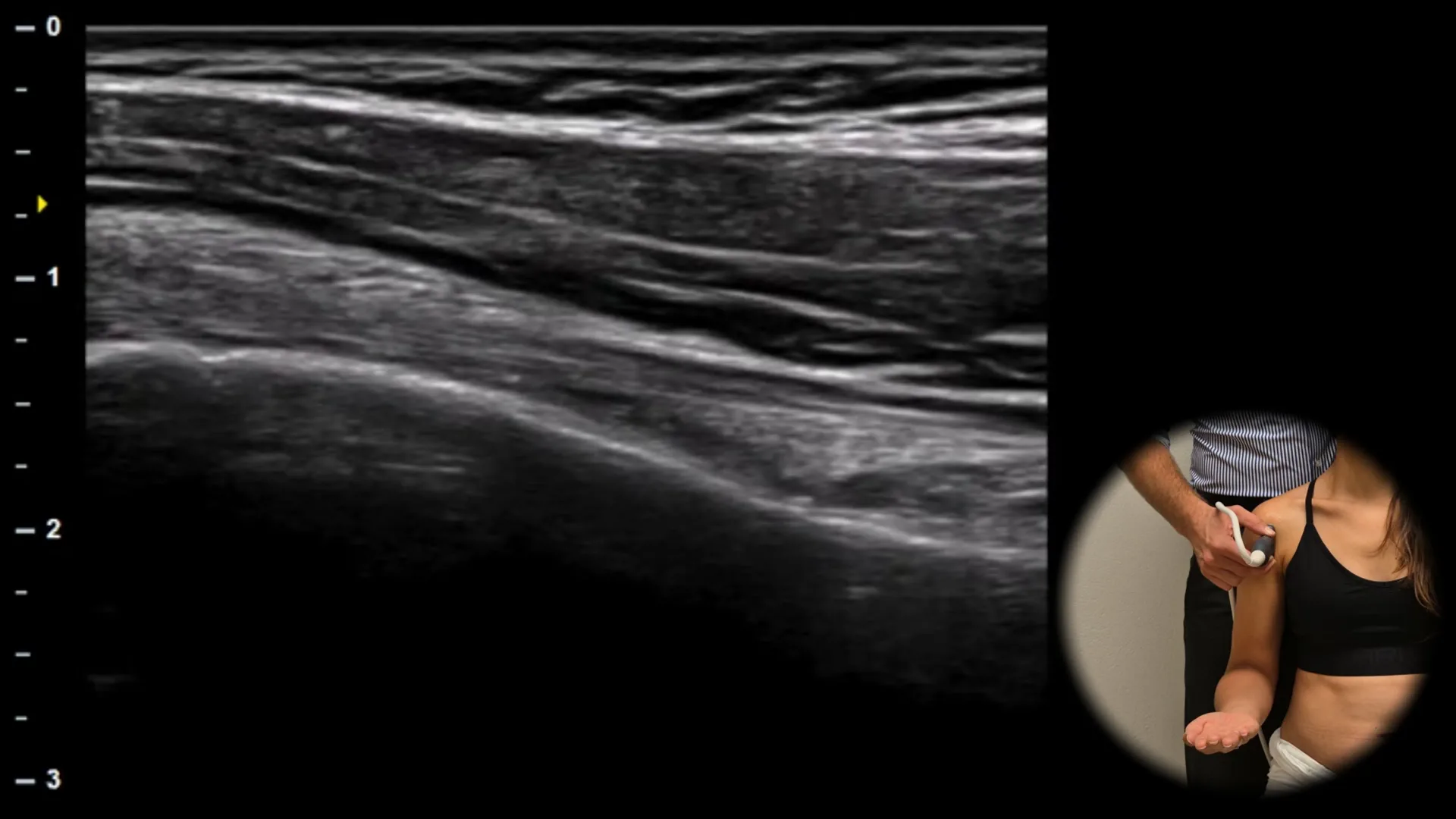
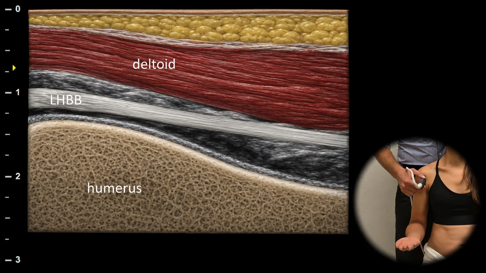
Figure 3. Ventral view, sagittal plane. b: tendon of the long head of m. biceps brachii
Longitudinal ultrasound section of the anterior shoulder, created by rotating the probe 90° from the transverse projection. The tendon of the long head of m. biceps brachii (b) is displayed in long axis in this plane as it courses through the intertubercular sulcus and shows the typical linear fibrillar appearance. This projection is essential for assessing tendon continuity and integrity and enables detection of pathological changes such as fluid around the tendon, tenosynovitis, or partial rupture.
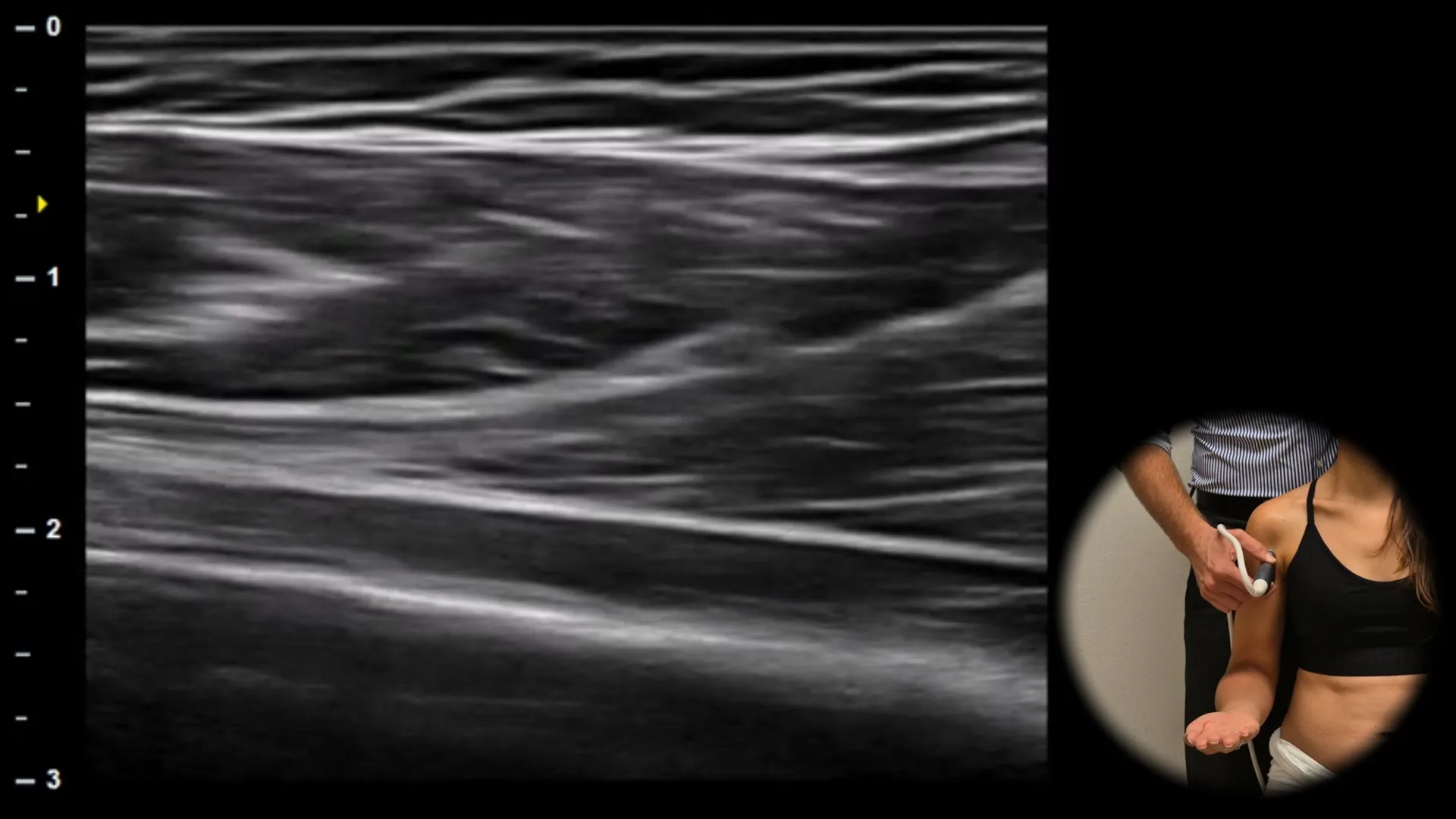
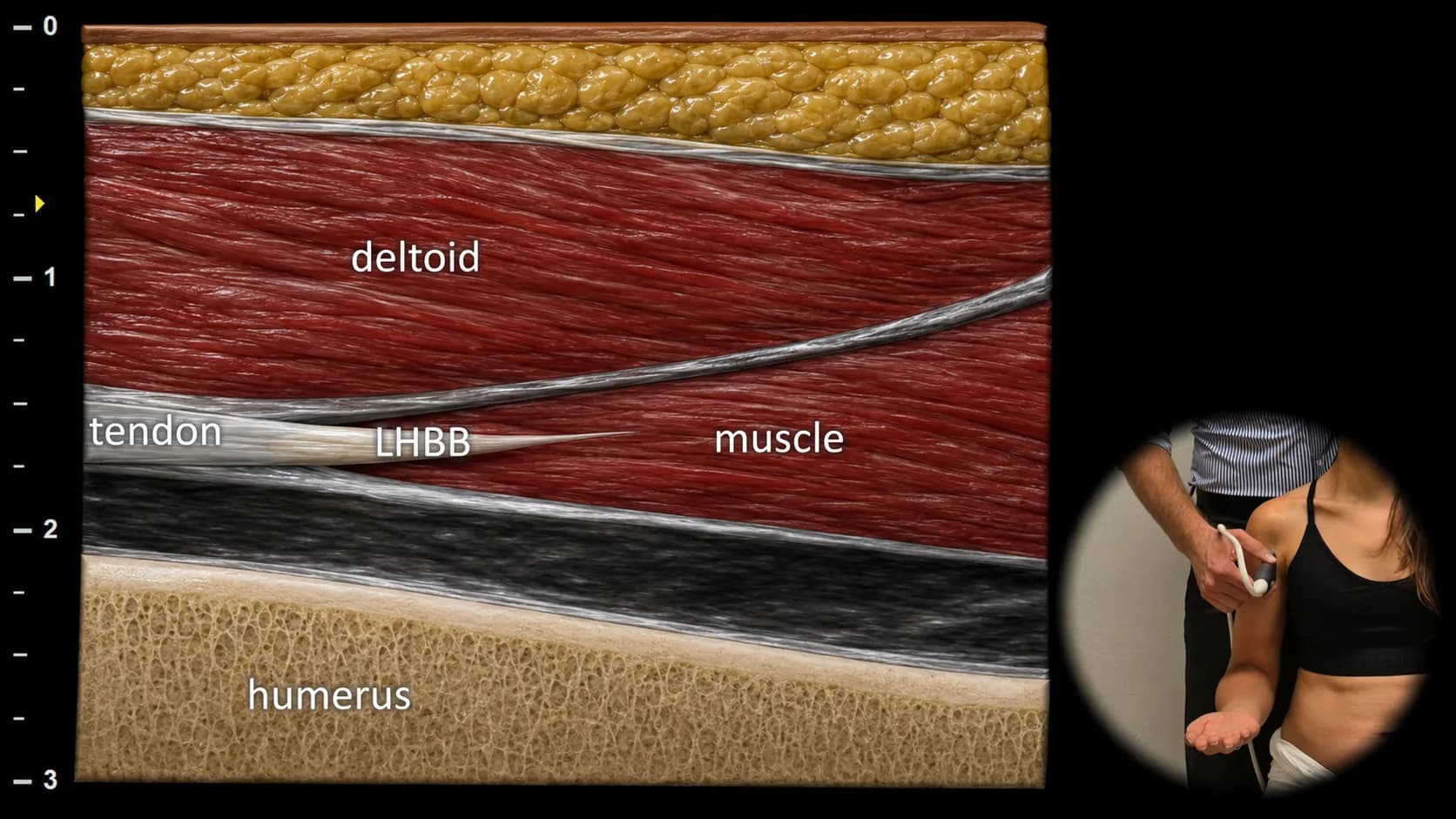
Figure 4. Ventral view, sagittal plane. LHBB: myotendinous junction of the long head of biceps brachii
Longitudinal ultrasound section of the anterior arm with more distal probe placement, showing the myotendinous junction area of the long head of biceps brachii (LHBB). In this projection, the transition from fibrillar tendon to hypoechoic muscle tissue is visible, corresponding to the transition from collagen fibers to muscle belly. This area represents a common site of injury and is important for detecting partial ruptures, tendinopathy, or muscle strain, especially in patients with anterior arm pain or following acute trauma.